Q55 - Filming Rotary Catalysis in F1-ATPase
F1 subunit (a peripheral membrane protein) of the ATP synthase catalyses ATP synthesis using proton motive force responsible for the rotation of F0 subunit (integral membrane protein complex) in one direction. F1 is composed of three alpha and three beta subunits arranged in alternating manner around a central shaft, the gamma subunit.
To study the rotation, Masasuke Yoshida and his team attached a fluorescently labelled actin filament to gamma and watched its movement.
Figure Q.55A Attachment of labelled actin filament to ATP synthase.
Rotating actin filaments were observed by an inverted fluorescence microscope after addition of 2 mM ATP into a chamber containing actin-tagged F1 complex immobilized on the bottom side as a mirror image formed on a camera. The time interval between images was 220 ms. A series of 12 images were taken and is shown in Fig. Q.55.
Figure Q.55B Sequential images of a rotating actin filament attached to the subunit in the F1 complex. The numbers indicate the shot images.
 Figure 1.
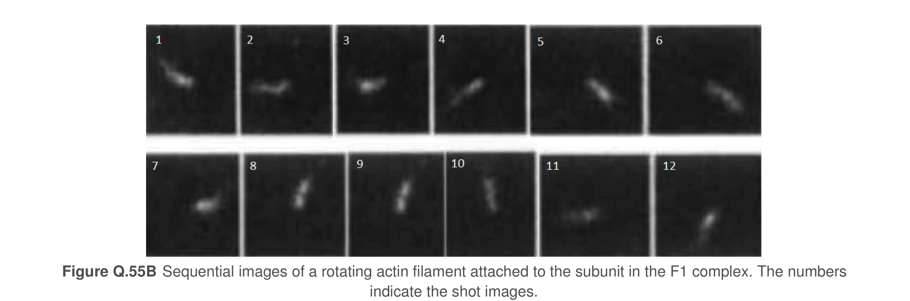 Figure 2.
Using the information and data, determine which of the statements are true or which are false.
Question reproduced from IBO 2016, Theoretical Paper B, licensed under CC BY-NC-SA 4.0 - attributed to the International Biology Olympiad. Open the full exam PDF · Community solutions (unofficial)