Q51 - Phage-Display Screen for Protein Kinase Inhibitors
A scientist prepared 3 essential components for high-throughput screens of protein kinase inhibitors. First, individual protein kinase genes are fused to the major capsid (head) gene of T7 phage. When expressed in bacteria, the fusion proteins are assembled into the phage capsid, with the kinases displayed on the outer surface. Second, an analog of ATP, which can bind to the ATP-binding pocket of the kinases, is attached to magnetic beads. Third, a bank of test compounds is prepared.
Figure Q.51 Screening potential inhibitors of protein kinases
To measure the ability of a test compound to bind a kinase, phage displaying a specific kinase is mixed with the magnetic beads in several wells of a 96-well plate. Then the test compound is added to individual wells over a range of different concentrations. The mixtures are incubated with gentle shaking for 1 hour at 25°C, the beads are pulled to the bottom with a strong magnet, and all the free (unbound) components are washed away. Finally, the remaining, attached phage are dissociated from the beads using an excess of the same ATP analog that is attached to the beads, and counted by measuring the number of plaques they form on a bacterial lawn on a Petri dish (Fig.Q.51).
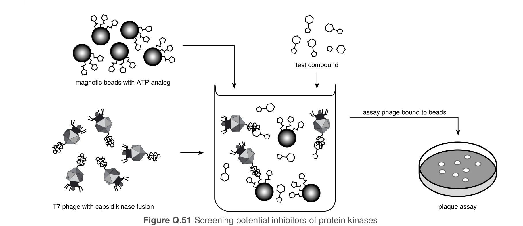 Figure 1.
Using the information and data, determine which of the statements are true or which are false.
Question reproduced from IBO 2016, Theoretical Paper B, licensed under CC BY-NC-SA 4.0 - attributed to the International Biology Olympiad. Open the full exam PDF · Community solutions (unofficial)