Q93 - Determining Insert Size and Orientation by Restriction Mapping
Mr. Trung cloned a coding sequence (CDS) of a gene into a vector and named the resulted plasmid as pVN2016. The CDS was inserted at the SacII recognition site which is located in the multi cloning site (MCS) region within the lacZ gene of the vector (Fig.Q.93A). The inserted CDS has a PstI restriction site located 0.8 kb upstream of its stop codon. To identify the size and direction of the inserted CDS, Mr. Trung digested this plasmid with different restriction enzymes, and the results of the digestions are shown in Fig.Q.93B.
Fig.Q.93 (A) A schematic map of the vector, showing the positions of the EcoRI, SpeI, and PstI recognition sites in the empty vector and the SacII cloning site. (B) A schematic electrophoresis gel of the digestion products from cutting pVN2016 with different single and combined restriction enzymes; M is a 1 kb DNA ladder.
Based on above data, indicate in the answer sheet if each of the following statements is true or false.
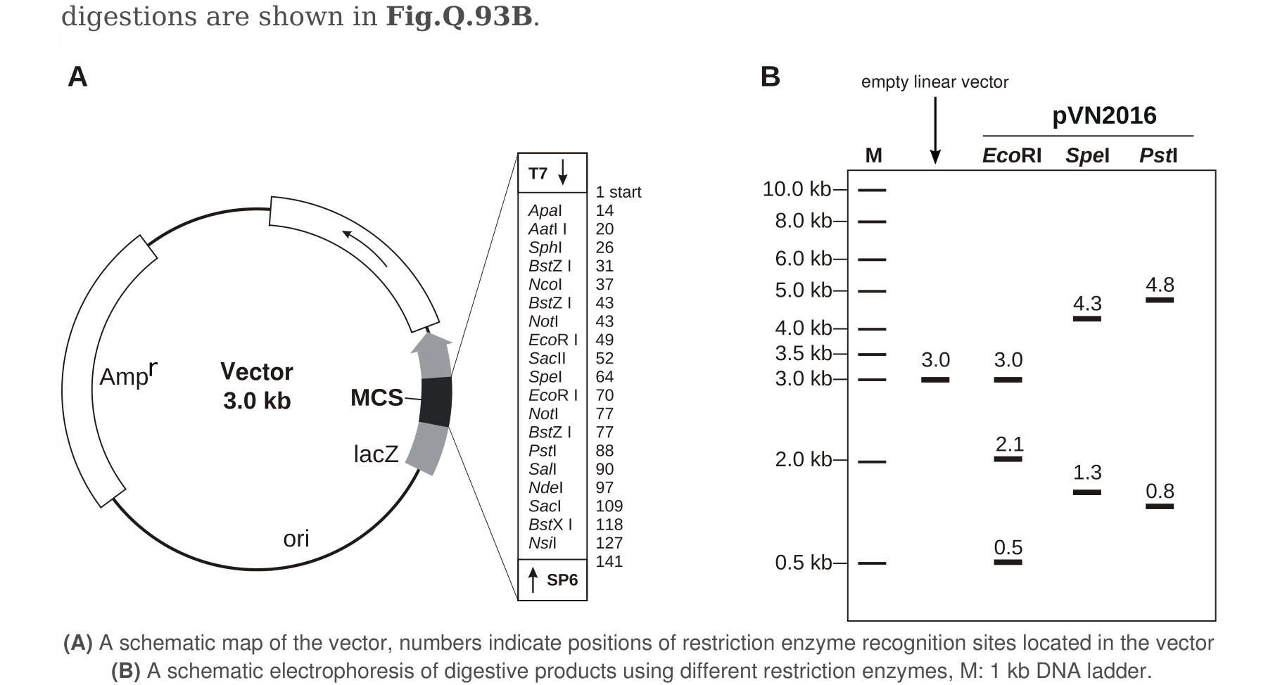 Figure 1.
Using the information and data, determine which of the statements are true or which are false.
Question reproduced from IBO 2016, Theoretical Paper B, licensed under CC BY-NC-SA 4.0 - attributed to the International Biology Olympiad. Open the full exam PDF · Community solutions (unofficial)