Q8 - Coordinating Enzymes at the E. coli Replication Fork
X-ray crystallography, invented at the Royal Institution, was used by Sir Francis Crick (1916-2004) and Rosalind Franklin (1920-1958) to discover the structure of DNA, and predict the mechanism of its replication. Building on their work, this is the current predicted structure of an E. coli DNA replication fork, which moves along DNA at 1000 bp/s. i = Tube shaped clamp, which pulls on one DNA strand. ii = Topoisomerase, which makes temporary cuts in one phosphodiester backbone. iii = Single-stranded DNA binding protein. iv = Different polymerases.
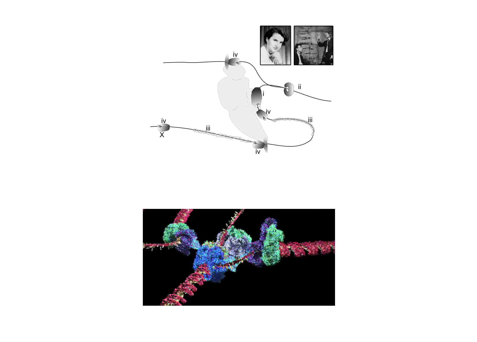
Using the information and data, determine which of the statements are true or which are false.
Question reproduced from IBO 2017, Theoretical Exam A, licensed under CC BY-NC-SA 4.0 - attributed to the International Biology Olympiad. Open the full exam PDF · Official answer key & worked solutions