DNA Structure & Replication
Overview
This page opens the Molecular Biology tier by covering how DNA is physically organised into a double helix, and the enzymatic machinery that copies it during S phase (see Cell Cycle, Mitosis & Meiosis). The chemistry of the nucleotides and phosphodiester backbone this page assumes is covered in Nucleotide & Nucleic Acid Chemistry; this page focuses on the higher-order double-helix structure and the replication process itself.
Key Concepts
The Watson-Crick double helix
DNA’s two strands run antiparallel (one 5′→3′, the other 3′→5′, running in opposite directions relative to each other) and wind around a common axis into a right-handed double helix, held together by two forces: hydrogen bonding between complementary base pairs (A–T, G–C, see Nucleotide & Nucleic Acid Chemistry for why only these pairings work chemically) running perpendicular to the helix axis, and base stacking (van der Waals/hydrophobic interactions between adjacent stacked bases) running parallel to it: base stacking actually contributes more to overall helix stability than hydrogen bonding alone, a frequently underappreciated point. The antiparallel arrangement is not incidental: it is a direct structural requirement for complementary base pairing to work geometrically at all, and it is the root cause of the leading/lagging strand asymmetry described below.

Why replication must be semiconservative
DNA replication produces two daughter molecules, each consisting of one original (template) strand and one newly synthesised strand: the semiconservative model, directly confirmed by the classic Meselson-Stahl density-gradient experiment (¹⁵N/¹⁴N isotope labelling), which distinguished it from two rival hypotheses: conservative replication (the original double helix stays fully intact, and an entirely new double helix is synthesised alongside it) and dispersive replication (both daughter molecules are a patchwork mixture of old and new DNA). Semiconservative replication is a direct mechanistic consequence of the fact that each template strand is individually read and directly base-paired against as the new strand is built, there is no step in the actual polymerisation mechanism that would produce either of the two rival patterns.

Origins of replication and the replication fork
Replication begins at specific origin of replication sequences, where initiator proteins recruit helicase, which unwinds the double helix, separating the two strands and creating a replication fork, the physical Y-shaped junction where unreplicated double-stranded DNA opens into two single-stranded templates. Single-strand binding proteins (SSBs) coat the exposed single strands, preventing them from re-annealing or forming secondary structures before replication machinery reaches them. Ahead of the fork, unwinding introduces mechanical torsional strain (supercoiling) into the still-double-stranded DNA, relieved by topoisomerases, which transiently cut and reseal the DNA backbone to release this strain.
DNA polymerase: capabilities and constraints
DNA polymerase synthesises new DNA by adding nucleotides complementary to the template strand, but is constrained by two fixed properties that shape everything downstream:
- It synthesises only in the 5′→3′ direction, reading the template 3′→5′.
- It cannot initiate a new strand from scratch: it can only extend an existing 3′-OH end, meaning every new strand (or fragment) requires a short pre-existing primer to begin from.
Primase, an RNA polymerase, solves the second constraint by synthesising a short RNA primer complementary to the template, providing the free 3′-OH end DNA polymerase requires to begin extension. This primer is later removed and replaced with DNA (see below).
Leading and lagging strands: the direct consequence of antiparallel geometry
Because the two template strands run in opposite directions, but DNA polymerase can only synthesise 5′→3′, the two new strands cannot both be synthesised continuously in the same direction the fork is opening:
- The leading strand is synthesised continuously, in the same direction the replication fork is moving, needing only a single primer at the origin.
- The lagging strand must be synthesised discontinuously, in short Okazaki fragments, each primed separately (new primer, new fragment) and each synthesised 5′→3′ but in the direction opposite to overall fork movement, because the template strand it’s copying runs the “wrong way” relative to the fork.
This asymmetry is entirely a geometric consequence of DNA polymerase’s fixed directionality meeting the antiparallel structure of the double helix, it is not an arbitrary or separately-evolved mechanism. On the lagging strand, RNA primers are subsequently removed and replaced with DNA (by a polymerase with 5′→3′ exonuclease activity, e.g. DNA polymerase I in bacteria) and the resulting nicks between adjacent Okazaki fragments are sealed by DNA ligase, forming a continuous strand.

Proofreading and fidelity
DNA polymerase’s overall low error rate comes from two layered mechanisms: selective nucleotide binding (correct Watson-Crick geometry is strongly preferred in the polymerase active site) and, when an incorrect nucleotide is nonetheless incorporated, 3′→5′ exonuclease proofreading activity, which detects the resulting geometric distortion, excises the mismatched nucleotide, and allows the polymerase to try again, a real-time editing function distinct from the post-replication mismatch repair covered in DNA Repair & Recombination, which catches errors that slip past proofreading entirely.
Comparative Structures
| Feature | Leading strand | Lagging strand |
|---|---|---|
| Synthesis pattern | Continuous | Discontinuous (Okazaki fragments) |
| Number of primers needed | One (at the origin) | Many (one per fragment) |
| Direction relative to fork movement | Same direction | Opposite direction (net) |
| Enzymes uniquely required | , | DNA ligase (fragment joining), 5′→3′ exonuclease (primer removal) |
| Replication model | Prediction | Outcome of Meselson-Stahl experiment |
|---|---|---|
| Conservative | One fully old + one fully new daughter molecule | Not observed |
| Semiconservative | Both daughters are old/new hybrids | Observed: confirmed model |
| Dispersive | Both daughters are patchworks of old/new DNA within each strand | Not observed |
Common Exam Questions
- “Why does the lagging strand require Okazaki fragments at all?”: the complete answer traces directly back to DNA polymerase’s fixed 5′→3′-only synthesis direction meeting the antiparallel template, a question asking “why” here wants the mechanistic chain, not just the definition of an Okazaki fragment.
- “What would you predict for the Meselson-Stahl experiment’s outcome under each replication model, and how did the observed result distinguish semiconservative from the alternatives?”, a frequently reconstructed classic-experiment reasoning question.
- Distinguishing polymerase proofreading (real-time, during synthesis, 3′→5′ exonuclease) from mismatch repair (post-synthesis, a separate pathway covered in DNA Repair & Recombination) is a common source of confusion worth explicitly separating.
Visual Reference
Interactive
- A replication fork step-through animation: helicase unwinding, SSB coating, primase laying a primer, and leading/lagging strand synthesis proceeding in real time, with Okazaki fragment formation and ligation visibly distinct from continuous leading-strand synthesis.
Static
(Static images are placed inline in Key Concepts above, next to the concept each one illustrates, rather than collected here. Note: the double-helix image has some visible annotation artifacts, see the inline note.)
Practice Challenge
Competition-sourced practice questions for this topic, graded by difficulty. Click the Solution tab to reveal each answer.
Theoretically, in human cells, the rate of DNA polymerase during replication is about 100 nucleotides per second. Assume that each copy of the human genome contains 3×10⁹ nucleotides and that both copies of the genome are identical.
A. What is the minimum number of origins of replication that such cells need to replicate their DNA every 12 hours? (Give your answer as an integer.)
B. On average, how many nucleotides are replicated from one origin within 12 hours? (Give your answer in millions of nucleotides to 2 decimal places.) ___ million nucleotides
The graph depicts the effect of different salt concentrations on the physical state of a bacterial DNA fragment. Select the most appropriate option that applies to this data.
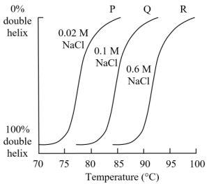
A. Increasing salt concentration increases the thermolability of the DNA strand.
B. Repulsion between like charges in DNA is more pronounced in condition P as compared to conditions Q and R.
C. Increasing salt concentration leads to increased hydrogen bonding between the DNA strands.
D. If one measures absorbance of the DNA strands at 260 nm, R is likely to show greatest absorbance at any temperature between 70-100°C as compared to P and Q.
Consider the following:
I) Bacteriophage genome, 10⁵ bp long, was fragmented to obtain unique (nonrepeating) fragments of 500 bp length each.
II) Bacterial DNA, 5×10⁶ bp long, was also fragmented to obtain unique (nonrepeating) fragments of 500 bp length each.
Equal quantity of each sample was denatured and allowed to re-associate separately under the same experimental conditions. During the re-association, the following parameters were tested:
i) S1 Nuclease digestion (specific for ssDNA) ii) Hypochromicity
At any point of time during the re-association, indicate whether each statement is True or False:
A. ‘I’ will show higher nuclease digestion value than ‘II’.
B. ‘II’ will show lower hypochromicity than ‘I’.
C. Both ‘I’ and ‘II’ will show equal reduction in absorbance at 260 nm but different nuclease digestion values.
D. Both ‘I’ and ‘II’ will show equal nuclease digestion values, but different absorbance values at 260 nm.
Practice Problems
1. A researcher performs the Meselson-Stahl experiment but observes, after one generation of replication in ¹⁴N medium following growth in ¹⁵N medium, a single intermediate-density band rather than two separate bands (one fully heavy, one fully light). Which replication model does this result support, and which two models does it rule out?
Show answer
This result supports the semiconservative model. A single intermediate-density band after one generation means every daughter molecule is a hybrid of one heavy (¹⁵N, old) and one light (¹⁴N, new) strand, exactly the semiconservative prediction. It rules out the conservative model (which predicts two distinct bands: one fully heavy, one fully light) and is also inconsistent with dispersive replication only once a second round of replication is examined (dispersive replication also gives one intermediate band after one generation, but predicts a shifting single band of decreasing density over further generations rather than two distinct bands, the true experiment required a second generation’s data to fully rule out dispersive replication, worth noting if a question specifically asks how conservative vs. dispersive were distinguished).
2. A mutant DNA polymerase completely lacks 3′→5′ exonuclease proofreading activity but is otherwise fully functional. Predict the effect on replication fidelity, and explain why this defect would not be expected to abolish replication entirely.
3. Explain why a primer, rather than DNA polymerase initiating synthesis directly, is required at the start of every Okazaki fragment, using DNA polymerase’s specific catalytic limitation.