Q20 - MRI Weighting and Comparative Organ Anatomy
The principle of magnetic resonance imaging (MRI) is based on the fact that the nuclei of certain elements align with the magnetic force when placed in a strong magnetic field. At the field strengths currently used in medical imaging, hydrogen nuclei (protons) in water molecules and lipids are responsible for producing anatomical images. If a radiofrequency pulse at the resonant frequency of hydrogen is applied, a proportion of the protons change alignment, flipping through a present angle, and rotate in phase with one another. Following this radiofrequency pulse, the protons realign (relax), they induce a signal which, although very weak, can be detected and localized by copper coils placed around the patient. An image representing the distribution of the hydrogen protons can be built up. The strength of the signal depends not only on proton density but also on two relaxation times, T1 and T2; T1 depends on the time the protons take to return to the axis of magnetic field, and T2 depends on the time the protons take to dephase. A T1-weighted image is one in which the contrast between tissues is due mainly to their T1 relaxation properties, while in a T2-weighted image the contrast is due to the T2 relaxation properties. T1-weighted image: Fat signal intensity > Water signal intensity T2-weighted image: Fat signal intensity < water signal intensity
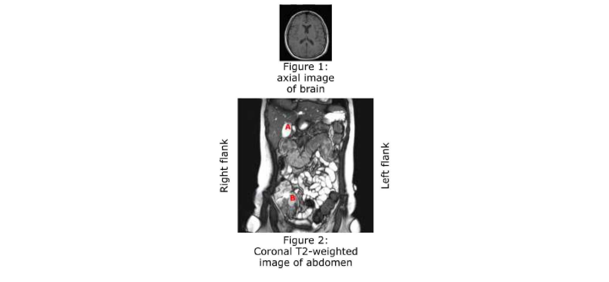 Figure 1.
Using the information and data, determine which of the statements are true or which are false.
Question reproduced from IBO 2018, Theoretical Paper 2, licensed under CC BY-NC-SA 4.0 - attributed to the International Biology Olympiad. Open the full exam PDF · Community solutions (unofficial)