Q6 - Identifying Cytoskeleton Types from Function and Morphology
Animal cells generally have three types of cytoskeletons: (1) microtubules, (2) actin filaments, and (3) intermediate filaments. Figure 1 shows the morphology of a cytoskeleton during the mitotic metaphase or in interphase. For each statement below A-E, indicate the corresponding type of cytoskeleton from (1) to (3), and the schematic diagram from (1) to (6) in Figure 1.
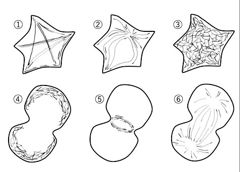 Figure 1.
Using the information and data, determine which of the statements are true or which are false.
A. They are entangled inside interphase cells and exist in a meshwork. They enhance the elasticity of cells and provide a mechanically supportive structure.
(3) intermediate filaments; diagram (3): A meshwork that gives cells mechanical elasticity and support, rather than driving movement or division, is the classic description of intermediate filaments, which form a rope-like, entangled network throughout the cytoplasm. In Figure 1 this tangled meshwork appearance corresponds to diagram (3).
B. It is called a stress fiber. It builds a support beam inside cells and works to maintain the shape of the cell in interphase.
(2) actin filaments; diagram (1): Stress fibers are a well-known, named structure built specifically from bundled actin filaments, which act as internal support beams anchoring the cell and maintaining its shape during interphase. In Figure 1 this corresponds to diagram (1), the crisscrossing bundle pattern typical of stress fibers.
C. This spindle-shaped structure is formed during cell division. It plays a role in separating replicated chromosomes accurately into daughter cells.
(1) microtubules; diagram (6): The spindle apparatus that pulls replicated chromosomes apart during division is built from microtubules, radiating from the two spindle poles toward the chromosomes at the cell's midline. In Figure 1 this corresponds to diagram (6), the two-cell radiating-fiber pattern typical of the mitotic spindle.
D. After chromosomal segregation, it forms a ring structure and mechanically separates two daughter cells.
(2) actin filaments; diagram (5): The contractile ring that pinches a dividing cell in two during cytokinesis, right after the chromosomes have separated, is built from actin (together with myosin motors). In Figure 1 this corresponds to diagram (5), showing the constriction at the junction between the two forming daughter cells.
E. Having radial distributions starting near the nucleus, the fibrous structure is assumed to have structural polarity or directionality.
(1) microtubules; diagram (2): A radial array of fibers emanating from a single organizing center near the nucleus is the signature interphase arrangement of microtubules, which grow outward from the centrosome with an intrinsic polarity (minus end at the center, plus end at the periphery). In Figure 1 this corresponds to diagram (2), the radiating star-shaped pattern.
Question reproduced from IBO 2020, Theoretical Exam 2, licensed under CC BY-NC-SA 4.0 - attributed to the International Biology Olympiad. Open the full exam PDF · Community solutions (unofficial)