Cytoskeleton & Motor Proteins
Overview
The cytoskeleton is not a static scaffold but a dynamic, constantly remodelling system of protein filaments providing mechanical support, intracellular transport tracks, and the machinery for cell shape change, motility, and division. Three distinct filament systems, differing in subunit protein, diameter, and mechanical properties, each specialise in a different structural role, and are read by three families of motor proteins that convert ATP hydrolysis into directed movement.
Key Concepts
Microfilaments (actin filaments)
The thinnest cytoskeletal filament (~7 nm diameter), built from polymerised globular actin subunits into a twisted double-helical filament. Actin filaments are polar: one end (the barbed/plus end) grows faster than the other (the pointed/minus end), and this polarity is functionally essential, both for directional motor movement (below) and for the treadmilling dynamics (net addition at the plus end, net loss at the minus end, giving apparent filament movement without net length change) that drive processes like the leading edge of a crawling cell. Microfilaments concentrate just under the plasma membrane (the cell cortex), providing mechanical support for cell shape, forming the core of microvilli, and, assembled with myosin, generating the contractile force of the cleavage furrow during cytokinesis and of muscle sarcomeres (see Cell Cycle, Mitosis & Meiosis for cytokinesis).

Microtubules
The thickest cytoskeletal filament (~25 nm diameter), built from α/β-tubulin dimers polymerising into a hollow tube of (typically) 13 protofilaments. Like actin, microtubules are polar (plus and minus ends), and undergo dynamic instability: individual microtubules stochastically switch between growth and rapid shrinkage phases, a property directly exploited during mitosis to search-and-capture chromosomes (see Cell Cycle, Mitosis & Meiosis). Microtubules radiate from a microtubule-organising centre (MTOC), the centrosome in most animal cells, with their minus ends typically anchored there and plus ends extending outward. Beyond mitotic spindle formation, microtubules form the structural core of cilia and flagella (the 9+2 axoneme arrangement) and serve as the principal long-distance intracellular transport highway.

Intermediate filaments
A structurally and functionally distinct third category (diameter ~10 nm, intermediate between the other two, hence the name), built from a diverse, tissue-specific family of fibrous proteins (keratins in epithelial cells, vimentin in mesenchymal cells, neurofilaments in neurons, lamins forming the nuclear lamina). Unlike actin and microtubules, intermediate filaments are non-polar (both ends structurally equivalent), are not tracks for motor proteins, and are considerably more mechanically stable/less dynamic: their role is purely structural, providing tensile strength and resisting mechanical stress, rather than participating in active transport or rapid remodelling. Lamins, an intermediate filament type unique to the nuclear interior, form a meshwork underlying the inner nuclear membrane that structurally supports the nucleus and must be depolymerised for nuclear envelope breakdown during mitosis.
Motor proteins: converting ATP hydrolysis into directed movement
Three motor protein families, each specific to one filament type and each moving unidirectionally along it:
- Myosin moves along actin filaments, generally toward the plus (barbed) end. Beyond its famous role in muscle contraction (myosin II thick filaments sliding past actin thin filaments: sarcomere shortening), myosin motors also transport vesicles and organelles along cortical actin.
- Kinesin moves along microtubules, generally toward the plus end (i.e., typically outward from the MTOC, toward the cell periphery): the principal motor for anterograde transport (e.g. moving vesicles/organelles from a neuron’s cell body out along the axon).
- Dynein moves along microtubules toward the minus end: retrograde transport (back toward the cell body/MTOC), and also the motor responsible for the coordinated beating of cilia and flagella, sliding adjacent microtubule doublets in the axoneme past each other.

All three convert the chemical energy of ATP hydrolysis into a cycle of conformational changes (a “power stroke”) that walks the motor along its filament track, and all three can carry cargo (vesicles, organelles, chromosomes via kinetochore-associated dynein) attached via adaptor proteins: the specific direction of net cargo movement in a cell is therefore determined by which motor is attached, not by the track alone.
Comparative Structures
| Filament type | Subunit protein | Diameter | Polarity | Motor protein(s) | Primary role |
|---|---|---|---|---|---|
| Microfilament | Actin | ~7 nm | Polar | Myosin | Cell cortex, contraction, cytokinesis |
| Microtubule | α/β-tubulin | ~25 nm | Polar | Kinesin (plus-end), dynein (minus-end) | Spindle, cilia/flagella, long-range transport |
| Intermediate filament | Tissue-specific (keratin, vimentin, lamin, etc.) | ~10 nm | Non-polar | None | Mechanical/tensile strength, nuclear lamina |
Common Exam Questions
- “Which cytoskeletal element is responsible for [structure/process]?” questions hinge on matching the specific filament to its role, cilia/flagella and the mitotic spindle are always microtubule-based; muscle contraction and the cell cortex are always actin-based; mechanical/tensile resistance and the nuclear lamina are always intermediate-filament-based.
- “A drug depolymerises microtubules, predict the effect on axonal transport and on mitosis”: tests whether you can trace a specific structural perturbation to both of a shared filament type’s downstream functions.
- Distinguishing kinesin (generally anterograde/outward) from dynein (generally retrograde/inward) direction is a frequent discriminator; note this is a generalisation about typical transport roles, not an absolute rule for every kinesin/dynein family member.
Visual Reference
Interactive
- A motor protein “walk” animator: select a motor (myosin/kinesin/dynein) and watch it step along its corresponding filament toward its characteristic end, carrying a cargo vesicle, makes the plus-end/minus-end direction rule visually concrete.
Static
(Static images are placed inline in Key Concepts above, next to the concept each one illustrates, rather than collected here.)
Practice Challenge
Competition-sourced practice questions for this topic, graded by difficulty. Click the Solution tab to reveal each answer.
Kinesin-5 motors are tetrameric motors that bind to anti-parallel microtubules and slide them apart. The microtubules in a mitotic spindle can be divided into three groups (1) kinetochore microtubules (ii) astral microtubules and (iii) interpolar microtubules as shown below.
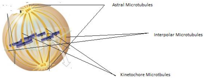
During mitosis, Kinesin-5 is most likely to be present on __________ microtubules and is expected to play a crucial role during _________. Choose the correct option to complete the given statement.
A. kinetochore and prophase B. interpolar and anaphaseC. astral and anaphaseD. interpolar and metaphase
Frog melanophores contain pigment granules that can either be dispersed throughout the cytoplasm, causing the cell to appear darker, or aggregated near the centre of the cell, causing it to appear lighter.
Pigment granules are transported along microtubules by motor proteins. The minus ends of microtubules are predominantly located near the cell centre, while the plus ends extend towards the cell periphery.
A researcher treats melanophores with different substances and measures pigment distribution.
(I) Which statement is most consistent with the role of microtubules in melanophore pigment transport?
A. Inhibition of microtubule polymerization would increase pigment aggregation at the cell centre because additional microtubules are formed with their minus ends facing outward.
B. A drug that stabilizes microtubules would necessarily prevent pigment granules from moving because motor proteins can only move along depolymerizing microtubules. C. If a motor protein transports pigment granules towards microtubule minus ends, increased activity of this motor would be expected to increase pigment aggregation near the centre of the melanophore.
D. If pigment granules move towards the plus ends of microtubules, depolymerization at the plus ends would increase the distance over which the granules can be transported towards the cell periphery.
(II) In a separate experiment, researchers measure the movement of pigment granules towards the centre of a melanophore following stimulation. The mean distance of pigment granules from the cell centre was measured at 10 time points:
| Time (s) | Mean distance from cell centre (μm) |
|---|---|
| 0 | 24.00 |
| 10 | 22.02 |
| 20 | 20.00 |
| 30 | 17.85 |
| 40 | 15.96 |
| 50 | 14.25 |
| 60 | 12.06 |
| 70 | 9.86 |
| 80 | 8.00 |
| 100 | 4.50 |
(a) Calculate the mean rate of movement of pigment towards the centre (in μm/s) :
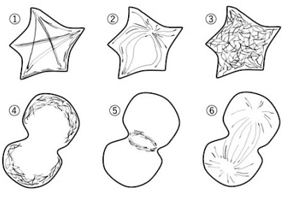
Animal cells generally have three types of cytoskeletons: (1) microtubules, (2) actin filaments, and (3) intermediate filaments. Figure 1 shows the morphology of a cytoskeleton during the mitotic metaphase or in interphase. For each statement below A-E, indicate the corresponding type of cytoskeleton from 1 to 3 in the first box, and the schematic diagram from 1 to 6 in Figure in the second box.
A. They are entangled inside interphase cells and exist in a meshwork. They enhance the elasticity of cells and provide a mechanically supportive structure. ()
B. It is called a stress fiber. It builds a support beam inside cells and works to maintain the shape of the cell in interphase.
C. This spindle-shaped structure is formed during cell division. It plays a role in separating replicated chromosomes accurately into daughter cells.
D. After chromosomal segregation, it forms a ring structure and mechanically separates two daughter cells.
E. Having radial distributions starting near the nucleus, the fibrous structure is assumed to have structural polarity or directionality.
| Statement | Cytoskeleton Type | Schematic Diagram |
|---|---|---|
| A | 3 | 3 |
| B | 2 | 1 |
| C | 1 | 6 |
| D | 2 | 5 |
| E | 1 | 2 |
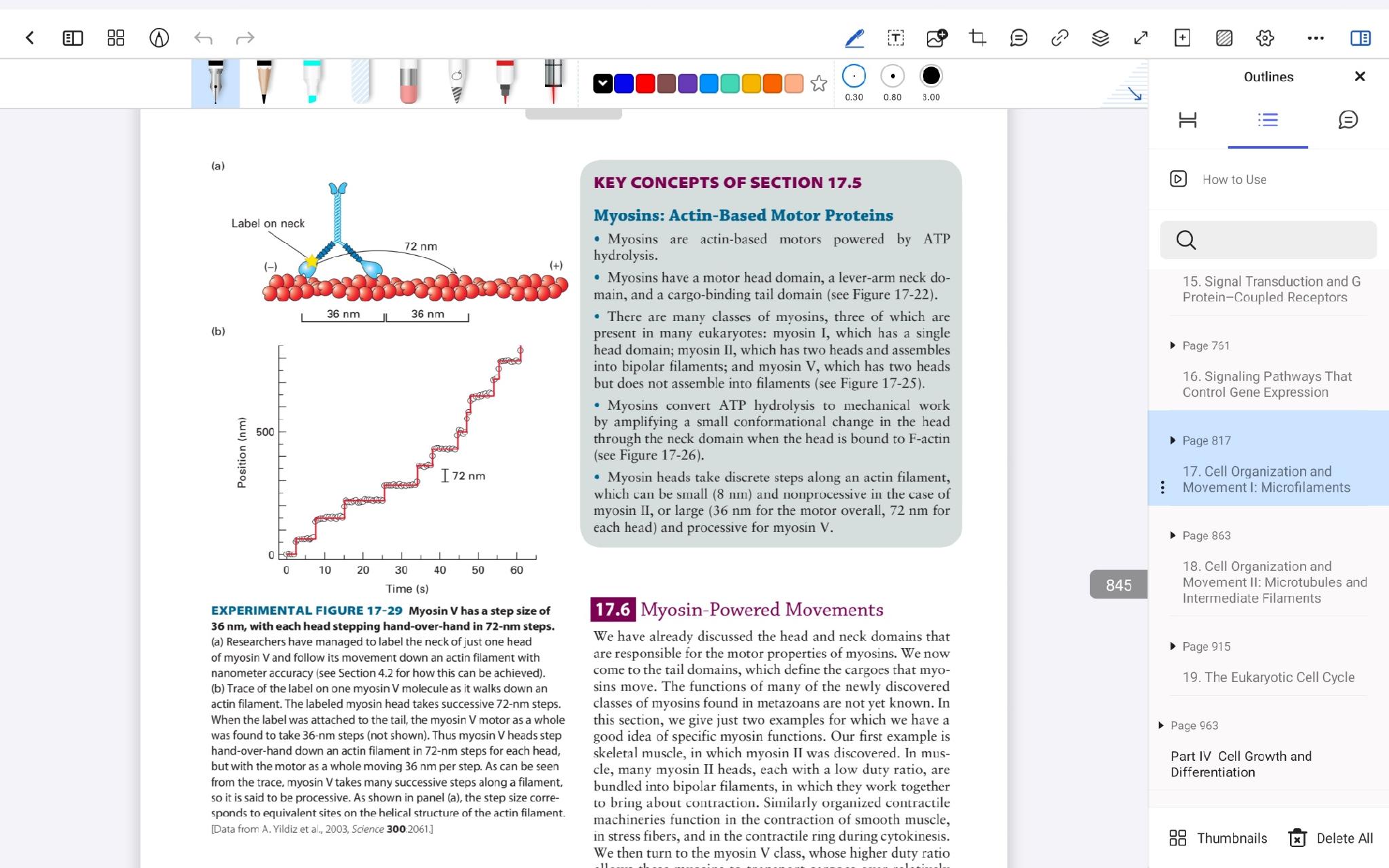
Myosin V is a motor protein that transports cargo along actin filaments. It has two motor heads, which bind to actin and move in an alternating hand-over-hand pattern. Researchers investigated the movement of individual myosin V molecules along immobilized actin filaments. To observe movement with high spatial resolution, they attached a fluorescent label to the neck region of one myosin V head. The actin filament was fixed in position, and the position of the fluorescent label was recorded repeatedly over time using fluorescence microscopy. The results of the experiment is given below in the figure. The researchers found that the labelled neck region did not move continuously. Instead, its position remained relatively constant for short periods before changing abruptly, producing a step-like trace similar to that shown in the figure. When one myosin head remains attached to the actin filament, the other head can detach and move past it before binding to a new site. The moving head may therefore travel a greater distance than the displacement measured for the labelled region of the entire motor.
Using the information provided and the figure, determine whether each statement is true or false:A. The step-like trace provides evidence that movement of myosin V along the actin filament occurs through discrete changes in position rather than at a constant continuous rate.
B. If the fluorescent label records a displacement of approximately 36 nm, each individual myosin head must also have moved exactly 36 nm during that step.
C. During hand-over-hand movement, one myosin head could move approximately 72 nm relative to its previous position while the overall position associated with the motor advances by approximately 36 nm.
D. Because myosin V contains two heads, an observed 36 nm displacement of the labelled motor can be interpreted as each head moving 18 nm during the same step.
Ans: A: T, B: F, C: T, D - F
Practice Problems
1. A cell is treated with a drug that specifically stabilises microtubules, preventing the dynamic instability (growth/shrinkage cycling) they normally undergo, without depolymerising them. Predict the effect on mitotic spindle function, and explain the mechanism.
Show answer
Mitosis is disrupted, likely arresting the cell at the point of chromosome capture/alignment. The mitotic spindle relies on dynamic instability for microtubules to “search” the cytoplasm by repeated growth and catastrophe until a plus end happens to encounter and stably attach to a kinetochore; artificially stabilising microtubules (preventing the shrinkage phase) removes this search-and-capture mechanism, so chromosomes fail to be efficiently captured and properly aligned at the metaphase plate.
2. A neuron’s axon shows normal outward (cell-body-to-synapse) vesicle transport but severely impaired return transport of used synaptic vesicle components back to the cell body. Which motor protein is most likely nonfunctional, and along which filament type does it operate?
3. Explain why intermediate filaments, despite providing critical mechanical support, are not used by the cell as tracks for directed cargo transport the way microtubules and microfilaments are: what structural property of intermediate filaments makes them unsuitable for this role?