Q13 - 3D Skin Substitute - Layered Tissue Engineering
For engineered tissue to survive in vivo it needs to be vascularized. Current approaches include incorporating adipose tissue-derived microvascular fragments, iPSC-derived endothelial cells, 3D bioprinting, and a layer-by-layer approach. The figure shows a multicellular 3D skin substitute assembled by interspersing three types of differentiated cells with thin layers of extracellular matrix film (fibronectin + gelatin), followed by several days of air-liquid interface culturing (Figure A). Figures B–D show staining outcomes: B - Masson’s trichrome (highlights collagen fibers), C - antibodies against laminin, D - antibodies against PECAM1 (an endothelial marker).
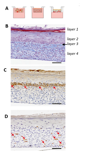 A: assembly of the 3D skin substitute. B: Masson’s trichrome staining, with layers 1–4 labelled. C: laminin staining (red arrows). D: PECAM1 staining (red arrows).
Using the information and figures, determine whether the following statements are true or false:
Question reproduced from IBO 2022, Theoretical Paper 1, licensed under CC BY-NC-SA 4.0 - attributed to the International Biology Olympiad. Open the full exam PDF · Community solutions (unofficial)