Q12 - iPSC/hESC Cardiomyocyte Grafts After Myocardial Infarction
Groundbreaking studies by Shinya Yamanaka’s group broke the long-standing dogma that differentiated adult cells cannot return to their embryonic state. The creation of induced pluripotent stem cells (iPSCs) offers hope that damaged organs can be repaired by a patient’s own cells. Multiple labs are working on therapies using patient-specific iPSC or immortalized human embryonic stem cell (hESC) lines - including pluripotent stem cell-derived cardiomyocyte grafts to re-muscularize scar tissue after myocardial infarction.
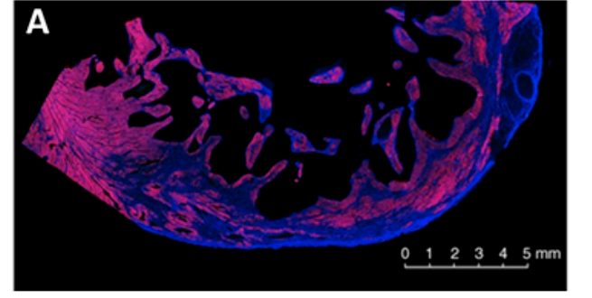 Fig. 1. Cross-section of the ventricular wall of an infarcted heart from a sham-treated monkey.
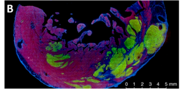 Fig. 2. Cross-section of the ventricular wall of an infarcted monkey heart with grafts of cardiac myocytes derived from hESC. The iPSCs were genetically modified to express green fluorescent protein (GFP).
Using the information and figures, determine whether the following statements are true or false:
Question reproduced from IBO 2022, Theoretical Paper 1, licensed under CC BY-NC-SA 4.0 - attributed to the International Biology Olympiad. Open the full exam PDF · Community solutions (unofficial)