Q81 - Blue Light, Melanopsin and the Circadian Phase-Shift Response
The human biological “internal clock” located in the suprachiasmatic nucleus (SCN) of the hypothalamus ensures the shift from sleep to waking state. To synchronize activity with the solar cycle, the SCN receives information from the light-sensing cells of the retina (rods, cones, ganglion cells), and reduces the synthesis of melatonin, which affects the activity of various processes. To assess the role of the retinal cells in regulating the sleep/wake cycle, a group of volunteers were kept in conditions of constant blue light (λmax = 460 nm) for 6.5h at night. Another group was kept in similar conditions but with green light (λmax = 555 nm). Cones have been shown to absorb green light, and retinal ganglion cells are capable of absorbing only blue light (λmax = 480 nm) with the help of melanopsin pigment. Melatonin levels of the subjects and the change in circadian rhythms - “phase shift” - were measured. The results are shown below.
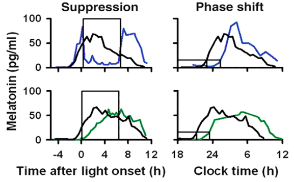 Figure 1. Melatonin levels during the experiment (left panels, exposure to light indicated by the black box) and the day after the experiment (right panels). Black curves are fluctuations in melatonin levels the day before the experiment. Blue curves (top panels) = blue light treatment (460nm), green curves (bottom panels) = green light treatment (555nm).
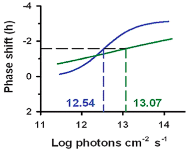 Figure 2. Dependence of the phase shift / change in circadian rhythm on the amount of light received. The dashed horizontal line shows half of the maximum daily rhythm shift, the dashed vertical lines show the proportions of light corresponding to those changes.
Using the information and data, determine which of the statements are true or which are false.
Question reproduced from IBO 2022, Theoretical Paper 2, licensed under CC BY-NC-SA 4.0 - attributed to the International Biology Olympiad. Open the full exam PDF · Community solutions (unofficial)