Q49 - Esophageal Doppler Monitoring
Esophageal Doppler Monitoring (EDM) is a minimally invasive method to measure blood flow in the aorta and assess cardiac output. A Doppler probe is inserted into the esophagus near the aorta. The principle behind the measurement is the detection of changes in frequency (Doppler shift) of ultrasound waves reflected by moving red blood cells. Using the Doppler effect, the probe calculates blood flow velocity from the equation Δf = 2vf₀cos(θ)/cₛ, where cos(θ) = √2/2, cₛ = 1540 m/s, and f₀ = 4 MHz.
The resulting waveform provides important data on heart function, such as stroke volume and cardiac output, which helps guide clinical decisions in real-time.
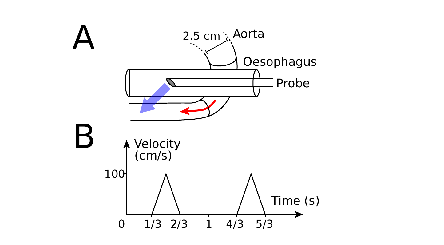 Figure 1. A Placement of the Doppler probe in the esophagus near the aorta to measure blood flow velocity (red arrow). B Doppler waveform showing linear blood velocity (cm/s) over time (s), with a peak systolic velocity of 100 cm/s. The area under the curve shows the distance traveled by blood.
On your answer sheet, indicate “T” for true statements and “F” for false ones.
Question reproduced from IBO 2024, Theoretical Exam Part A, licensed under CC BY-NC-SA 4.0 - attributed to the International Biology Olympiad. Open the full exam PDF · Community solutions (unofficial)