Q9 - ATAD1 Mitochondrial ATPase (Cryo-EM Study)
Cryo-EM is a modern structural study technique that works by averaging multiple low-quality electron micrographs; cryo-EM generates high-quality structural maps. It offers flexibility by accommodating various molecular sizes and capturing multiple conformations. The structure and mechanism of ATAD1, a mitochondrial homohexamer protein, were resolved using cryo-EM. The protein’s function was then determined using the structural data and fluorescent assays.
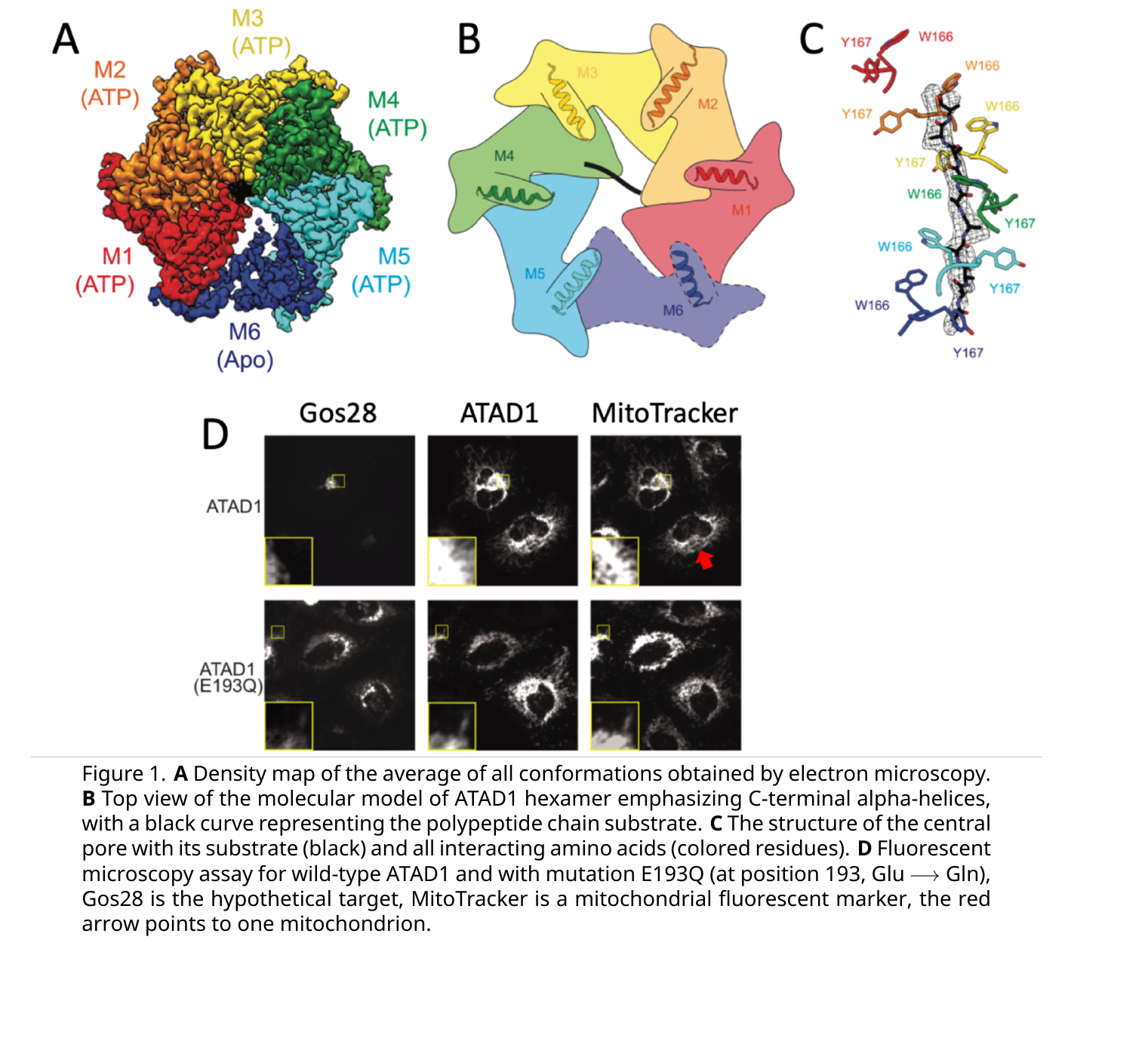 Figure 1. A Density map of the average of all conformations obtained by electron microscopy. B Top view of the molecular model of ATAD1 hexamer emphasizing C-terminal alpha-helices, with a black curve representing the polypeptide chain substrate. C The structure of the central pore with its substrate (black) and all interacting amino acids (colored residues). D Fluorescent microscopy assay for wild-type ATAD1 and with mutation E193Q (at position 193, Glu → Gln), Gos28 is the hypothetical target, MitoTracker is a mitochondrial fluorescent marker, the red arrow points to one mitochondrion.
On your answer sheet, indicate “T” for true statements and “F” for false ones.
Question reproduced from IBO 2024, Theoretical Exam Part A, licensed under CC BY-NC-SA 4.0 - attributed to the International Biology Olympiad. Open the full exam PDF · Community solutions (unofficial)