Protein Structure, Folding & Function
Overview
A protein’s function is inseparable from its shape, and its shape is built in four hierarchical levels: primary, secondary, tertiary, and quaternary structure, each stabilised by a different balance of covalent and non-covalent forces. This page covers that hierarchy, the model system for allosteric regulation (hemoglobin), how cells prevent and correct misfolding, and how they destroy proteins deliberately. Notably, protein fold is more evolutionarily conserved than amino acid sequence. Distantly related proteins with very different sequences often adopt near-identical folds, because the geometric and energetic constraints on a stable fold are stricter than the constraints on any single residue.
Key Concepts
The four levels of structure
Primary structure is simply the linear amino acid sequence, held together by peptide bonds (and, for cross-chain or cross-loop connections, disulfide bonds between cysteines).
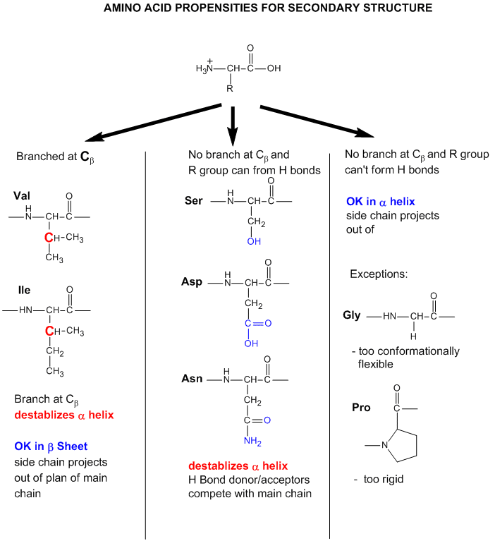
Secondary structure is a local, repeating backbone conformation stabilised almost entirely by hydrogen bonds between the carbonyl oxygen and amide hydrogen of the polypeptide backbone, side chains are not directly involved. Two dominant motifs:
- α-helix (one turn = 3.6 residues). High-propensity residues: Ala, Glu, Leu, Met, Lys (“MALEK”), small enough to pack tightly, or able to form stabilising salt bridges (Glu, Lys), with no steric interference with the coil. Helix breakers: Proline (its side chain cyclises onto the backbone nitrogen, removing the amide hydrogen the helix needs and imposing a rigid kink) and Glycine (too flexible, entropically costly to lock into one rigid helical conformation).
- β-sheet (≈180° turn per 4 residues). High-propensity residues: bulky aromatics (Trp, Tyr, Phe) and β-branched residues (Val, Ile, Thr). Their bulk disfavours the tight coiling of a helix and favours the more open, extended sheet geometry. Sheet breakers: Proline (same rigidity argument) and bulky charged residues (Glu, Lys) that clash sterically/electrostatically in the tightly packed strand.
- β-turn: a third, shorter motif connecting two antiparallel β-strands.

 Source: Unknown
A useful rule of thumb: secondary-structure hydrogen bonding is local, roughly n to n+3/n+4, only nearby residues interact directly.
Supersecondary structure (motifs) combine a small number of secondary elements in a recognisable pattern, α-α-α, α-β-α, β-β-β, without yet forming a full folded domain.

Tertiary structure is the overall 3D fold of a single polypeptide, formed by packing secondary elements into compact domains. It is stabilised by, in rough order of contribution:
- Hydrophobic interactions: the dominant driving force; nonpolar side chains bury in the interior to avoid water, polar side chains stay solvent-exposed.
- Hydrogen bonds between polar side chains, or side chain–backbone, individually weak but numerous and directional.
- Ionic bonds (salt bridges) between oppositely charged side chains (e.g. Lys⁺···Asp⁻).
- Disulfide bonds, the only covalent tertiary-level bond, cross-linking two cysteine sulfurs.
- Van der Waals forces: weak, short-range, but numerous in a densely packed hydrophobic core.

Because most of these forces are non-covalent, tertiary structure is fragile: heat, extreme pH, or heavy metals disrupt them, causing denaturation, loss of function without necessarily breaking the primary sequence.
Quaternary structure arises when multiple independently folded (tertiary) subunits associate, often via the same non-covalent forces listed above, into one functional multimeric complex.

Hemoglobin: the model system for allostery and cooperativity
Allostery is regulation of a protein’s activity by a ligand binding somewhere other than the active/functional site, via an induced conformational change. Hemoglobin (Hb), a tetramer of two α- and two β-globin chains, is the canonical teaching example.
T (tense) vs. R (relaxed) state: deoxy-Hb sits in the T state, held by 8 inter-subunit salt bridges, with low O₂ affinity. When O₂ binds one heme iron, the iron (previously pulled slightly out of the porphyrin plane by the proximal histidine) moves into the plane, tugging the attached helix and breaking salt bridges, a conformational change that propagates cooperatively across all four subunits, flipping the whole tetramer to the R state with high O₂ affinity.

Cooperativity means O₂ binding at one subunit raises affinity at the others, producing a sigmoidal binding curve (vs. the hyperbolic curve of monomeric myoglobin, which has no cooperativity):
For Hb, the Hill coefficient n_H ≈ 2.8 (out of a theoretical maximum of 4 for perfect cooperativity); for myoglobin, n_H = 1. You do not need to derive this equation for most olympiad purposes, understand what n_H means (degree of cooperativity) and its bounds.

Heterotropic effectors: ligands other than O₂ that shift Hb’s affinity:
| Effector | Effect on O₂ affinity | Mechanism |
|---|---|---|
| 2,3-bisphosphoglycerate (2,3-BPG) | Decreases | Binds the central cavity of the T state, stabilising it |
| CO₂ | Decreases | Forms carbamino-Hb; also lowers pH |
| H⁺ (low pH, the Bohr effect) | Decreases | Protonates β-chain His HC3, stabilising T state |
| CO | Increases (pathologically) | Binds the same site as O₂ with >200× affinity; shifts remaining subunits toward R, reducing effective cooperativity and causing hypoxia despite normal Hb saturation |
At the pO₂ found in respiring tissue (~40 mmHg), myoglobin (with its hyperbolic, high-affinity curve) retains far more bound O₂ than hemoglobin, which is exactly the point of the two proteins having different curve shapes: Hb is built to release O₂ readily in tissue, Mb to hold onto it as an intracellular O₂ store.

Chaperones and protein folding
Anfinsen’s dogma: a protein’s final fold is fully encoded in its primary sequence, chaperones do not dictate the fold. What they do is prevent off-pathway aggregation, by transiently shielding exposed hydrophobic patches during and after translation, before folding completes.
GroEL/GroES (Hsp60 family) cycle: (1) the unfolded protein enters the GroEL barrel, whose inner surface is initially hydrophobic; (2) ATP binding flips the inner surface hydrophilic, creating a protected folding chamber; (3) the GroES cap closes over the chamber; (4) ATP hydrolysis (~10 s later) releases the protein, correctly or incorrectly folded; (5) incorrectly folded protein re-enters the cycle.
Hsp70 (DnaK in bacteria) binds extended peptide co-translationally, assisted by co-chaperones DnaJ (Hsp40) and the nucleotide-exchange factor GrpE.
Misfolding diseases
| Disease | Protein | Misfolding event |
|---|---|---|
| Sickle cell anaemia | β-globin (HbS, Glu6Val) | The substituted valine creates a hydrophobic surface patch; deoxy-HbS polymerises into rigid fibres |
| Cystic fibrosis | CFTR (most commonly ΔF508) | Misfolded CFTR is retained in the ER and degraded by ERAD before ever reaching the plasma membrane |
| Prion disease (CJD, scrapie) | PrP | Native α-helix-rich PrPᶜ converts to β-sheet-rich PrPˢᶜ; PrPˢᶜ templates the same conversion in native PrPᶜ molecules it contacts, a self-propagating misfold requiring no nucleic acid |
| Alzheimer’s disease | Amyloid-β / Tau | Aβ aggregates into cross-β amyloid plaques; Tau forms intracellular neurofibrillary tangles |
| Parkinson’s disease | α-synuclein | Aggregates into Lewy bodies |
Prion disease is a favourite exam topic precisely because it breaks the central dogma’s usual information flow: heritable, infectious biological information is propagated by protein conformation alone, with no nucleic acid template. This is also why prions resist UV/nuclease treatment (which target nucleic acids) and require harsh treatment (autoclaving with NaOH) for inactivation.
The ubiquitin-proteasome system
Beyond phosphorylation and glycosylation, the ubiquitin-proteasome system (UPS) is the cell’s primary route for regulated, targeted protein destruction. Ubiquitin (Ub), a 76-residue protein, is attached to a target lysine via an isopeptide bond in a three-enzyme cascade:
$$ \text{E1 (Ub-activating)} \rightarrow \text{E2 (Ub-conjugating)} \rightarrow \text{E3 (Ub-ligase, provides substrate specificity)} $$
The linkage type determines the outcome:
- Lys48-linked polyubiquitination → targets the protein for proteasomal degradation
- Lys63-linked polyubiquitination → non-degradative signal (DNA repair, endosomal sorting)
- Monoubiquitination → histone regulation, membrane protein trafficking
The 26S proteasome is a 20S catalytic barrel (with chymotrypsin-like, trypsin-like, and caspase-like protease activities) capped by two ATP-dependent 19S regulatory particles, which recognise polyubiquitin chains, unfold the substrate, and thread it into the barrel. This system is the drug target of bortezomib, a proteasome inhibitor used in multiple myeloma.
Fibrous vs. globular proteins
Structural role correlates strongly with secondary-structure composition: α-keratin is helix-rich with disulfide cross-links, giving toughness; silk fibroin is β-sheet-rich, giving flexibility over tensile toughness; collagen forms a unique triple helix (distinct from the α-helix), giving very high tensile strength. Globular proteins (e.g. hemoglobin) fold compactly with a hydrophobic core, favouring solubility and functional flexibility (conformational switching, as in the T↔R transition above) over mechanical strength.
Comparative Structures
| Feature | α-Helix | β-Sheet |
|---|---|---|
| H-bond pattern | Intrachain, local (i to i+4) | Interchain or distant intrachain, extended |
| Residues per turn/repeat | 3.6 residues/turn | ~2 residues per repeat unit, sheet is a zig-zag |
| High-propensity residues | Ala, Glu, Leu, Met, Lys | Trp, Tyr, Phe, Val, Ile, Thr |
| Breakers | Pro, Gly | Pro, bulky charged residues (Glu, Lys) |
| Typical role | Membrane-spanning segments, structural coils (keratin) | Structural sheets (fibroin), extended binding surfaces |
Common Exam Questions
- “Explain why proline and glycine disrupt α-helices”: always give the chemical reason (cyclic side chain / no amide H for Pro; excess backbone flexibility for Gly), not just “they’re different.”
- “At tissue pO₂, which protein (hemoglobin or myoglobin) holds more oxygen, and why does this matter physiologically?” This tests whether you understand why the two curve shapes exist, not just that they differ.
- Distinguishing whether a stabilising interaction is covalent (disulfide bond only) vs. non-covalent (everything else in tertiary structure) is a common trap in “which bond breaks first on denaturation” questions.
- Prion disease questions often test the “no nucleic acid required” concept specifically, watch for answer choices that assume all heritable/infectious agents must involve DNA or RNA.
Visual Reference
Interactive
- A clickable hemoglobin T-state/R-state toggle: view the tetramer, click to trigger the conformational shift, watch the salt bridges break and reform.
Static
(Static images are placed inline in Key Concepts above, next to the concept each one illustrates, rather than collected here. One item (a primary-structure diagram showing a peptide bond chain with a disulfide cross-link) has no sourced image yet: Still needs sourcing.)
Practice Challenge
Competition-sourced practice questions for this topic, graded by difficulty. Click the Solution tab to reveal each answer.
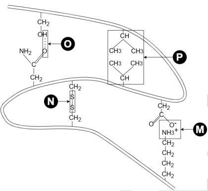
A portion of the tertiary structure of a protein is represented below.
M, N, O and P respectively represent:A . ionic bond; disulphide bridge; hydrogen bond and hydrophobic interactionsB . hydrophobic interactions; ionic bonds; disulphide bridge and hydrogen bondC . hydrogen bond; disulphide bridge; ionic bond and hydrogen bondD . ionic bond; disulphide bridge; hydrophobic interaction and hydrogen bond
Which of the following bonds/interactions involved in folding a protein into its proper 3D shape is INCORRECTLY matched with its description?
A. Hydrogen bond: a bond formed by the attraction of a hydrogen atom to an electronegative atomB. Hydrophobic effect: the attraction between a hydrophobic molecule and a hydrophilic moleculeC. Electrostatic interaction (ionic bond): the bond between a positively charged ion and a negatively charged ionD. Van der Waals force: an attractive force that occurs due to resonating dipole moment when atoms or groups of atoms are very close together
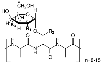
Antifreeze glycoproteins (AFGPs) possess the ability to inhibit the formation of ice and are therefore essential to the survival of many marine teleost fishes that routinely encounter sub-zero temperatures. A typical AFGP consists of repeating tripeptide units, the alanyl-threonyl-alanyl (Ala-Thr-Ala)n unit connected to a disaccharide through a glycosidic bond at the second hydroxyl group of the threonine residue. To identify chemical groups which affect antifreeze activities of this glycoprotein, scientists synthesized numerous AFGP analogues by modifying both the structure of the sugar moieties and the peptide by replacing three groups R1, R2, R3 as shown in Figure with different chemical groups and recorded the antifreeze activity.
The results of the study are shown in the following table.
| R1 | R2 | R3 | Antifreeze Activity |
|---|---|---|---|
| HO | CH3 | Galactosyl | No |
| N-Acetyl | CH3 | Galactosyl | Yes |
| N-Acetyl | H | Galactosyl | No |
| N-Acetyl | CH3 | H | Yes |
| O-Acetyl | CH3 | H | No |
| N-Acetyl | CH3 | Galactosyl-Galactosyl | No |
Indicate in the Answer Sheet if each of the following statements is True or False: A. A disaccharide bound to the threonine residue is required for antifreeze activity. B. A mutant that has threonine residues replaced with serine residues reduces antifreeze activities. C. N-acetyl group at the C-2 position is required for antifreeze activity. D. Different numbers of repetitive motifs in AFGP genes amongst closely related species might have been caused by DNA polymerase inaccuracy.
Practice Problems
1. Plot (conceptually) the O₂-binding curves of hemoglobin and myoglobin on the same axes. At pO₂ = 40 mmHg (venous tissue), which protein retains more O₂, and why is this the physiologically correct behaviour for each protein’s role?
Show answer
Myoglobin’s hyperbolic curve sits to the left of (higher affinity than) hemoglobin’s sigmoidal curve across the whole range, so at pO₂ = 40 mmHg myoglobin is still highly saturated while hemoglobin has released a substantial fraction of its bound O₂. This is functionally correct: myoglobin’s job is to store O₂ inside muscle cells and only release it under extreme local hypoxia (heavy exertion); hemoglobin’s job is to transport O₂ and unload a large, tunable fraction of it in respiring tissue: the sigmoidal curve, shifted further right by 2,3-BPG, CO₂, and low pH exactly where those signals of active metabolism are highest, makes this unloading responsive to local tissue demand.
2. A missense mutation converts a helix-interior alanine to proline in an otherwise stable α-helical domain. Predict the structural consequence, and explain the mechanism.
Show answer
Inserting proline mid-helix introduces a rigid kink and removes an amide hydrogen the helix’s hydrogen-bonding pattern requires at that position, locally disrupting or terminating the helix. The mutation is likely destabilising unless the position happens to sit at a natural turn/kink already present in the native structure.
3. Two proteins have identical Lys48-linked polyubiquitin chains but different fates: one is degraded within minutes, the other persists for hours despite similar chain length. Propose two non-ubiquitin-related explanations.
4. Explain, in mechanistic terms, why CO poisoning causes tissue hypoxia even when overall Hb-bound oxygen content may not be far below normal.