Q5 - Fluorescence Quenching of Tryptophan Residues
Enzyme activity usually correlates with its conformational flexibility, such that higher flexibility (lower rigidity) is usually accompanied with higher activity. Tryptophan residues which emit fluorescence are most commonly located within the nonpolar interior environment of the proteins. An excellent way to experimentally determine the exposure of tryptophan residues to solution is by measuring the quenching (decrease) of their fluorescence. The effect of mutations on the accessibility of tryptophan residues can be studied by measuring amount of fluorescence quenching by potassium iodide (KI). Iodide ions selectively quench fluorescence emitted by exposed tryptophan residues. In the experiment whose results are presented below, fluorescence quenching on equal amounts of three mutated forms of an enzyme (mutant forms 1, 2, and 3) was measured after addition of various concentrations of KI (0-0.6 M). Excitation and emission wavelengths used were specific for tryptophan. Quenching data were analysed in terms of the Stern-Volmer constant, KSV, which can be calculated from the ratio of the unquenched and the quenched fluorescence intensities, Fo/F, using the relationship F0/F = 1 + KSV[Q], where [Q] is the molar concentration of the quencher.
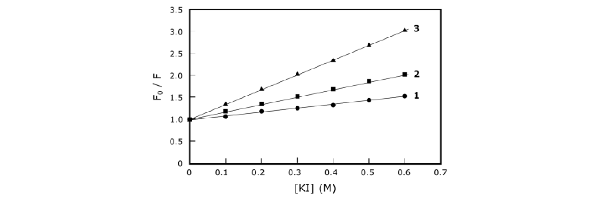 Figure 1.
Using the information and data, determine which of the statements are true or which are false.
Question reproduced from IBO 2018, Theoretical Paper 1, licensed under CC BY-NC-SA 4.0 - attributed to the International Biology Olympiad. Open the full exam PDF · Community solutions (unofficial)