Q3 - Flow Cytometry of Mouse Blood
Flow cytometry is a method for analyzing multiple cells in suspension. Cells pass through a microfluidic system, where they are irradiated by lasers. Laser light photons are partially scattered by cells and partially absorbed by fluorophores (fluorescent molecules). Excited fluorophores then emit secondary photons at longer wavelengths.
Fluorophores are typically small organic molecules with conjugated double bonds.
- Low angle scattering is measured by a forward scattering (FSC) detector.
- High angle scattering is measured by a side scattering (SSC) detector.
- Fluorescence is measured by wavelength-specific detectors.
FSC positively correlates with cell size, while SSC positively correlates with cell-surface “texture” and “granularity,” including protrusions, pockets and underlying vesicles.
The results of measurement are typically represented on dot-plot diagrams. Each dot represents a single cell, with the density of dots in an area indicated by color (red is high density, dark blue is low density).
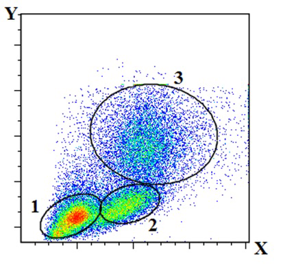 Flow cytometry analysis of mouse blood pretreated with red blood cell lysis buffer. X = FSC, Y = SSC. An FSC-threshold was used to exclude particles with very low FSC (e.g. debris).
Indicate whether the following statements are true or false:
Question reproduced from IBO 2023, Theoretical Paper 1, licensed under CC BY-NC-SA 4.0 - attributed to the International Biology Olympiad. Open the full exam PDF