Biomolecular Analytical Techniques
Overview
Olympiad practical exams routinely test technique choice, given a purification or analysis goal, pick and justify the right method, more than technique trivia. This page covers the core protein-analysis toolkit: electrophoresis (how it separates, and what “native” vs. “denaturing” actually changes), the three major chromatography modes and what property each separates on, spectrophotometric quantification, and how detergent choice determines whether a membrane protein extraction preserves native function. For nucleic-acid-specific techniques (blotting, PCR, sequencing), see Recombinant DNA & Biotechnology Techniques. This page is protein-focused.
Key Concepts
SDS-PAGE: denaturing electrophoresis by size alone
SDS-PAGE (sodium dodecyl sulfate–polyacrylamide gel electrophoresis) separates proteins almost purely by molecular size, not native charge or shape. SDS is an anionic detergent that (1) denatures proteins, unfolding tertiary/quaternary structure, and (2) coats the unfolded polypeptide with a uniform negative charge roughly proportional to its length. This second effect is what makes size, not intrinsic charge, the dominant determinant of migration rate. Samples are typically also treated with a reducing agent (e.g. β-mercaptoethanol or DTT) to break disulfide bonds, fully separating multi-chain or disulfide-linked proteins into their individual polypeptides. Smaller polypeptides migrate faster through the polyacrylamide mesh toward the anode; comparing migration distance to a ladder of known-size marker proteins gives an estimated molecular weight.

Native PAGE: preserving structure and function
Native PAGE omits SDS and reducing agent, so proteins retain their folded, native conformation, native charge, and any quaternary assemblies (e.g. a tetramer runs as one band, not four). Separation now depends on a combination of size, shape, and intrinsic charge, messier to interpret for pure size determination, but essential when the goal is preserving activity (e.g. a subsequent in-gel enzyme activity assay, which requires the enzyme to still be folded and catalytically competent).
Agarose gel electrophoresis: the nucleic acid analogue
For nucleic acids, agarose gel electrophoresis plays a directly analogous role to SDS-PAGE: DNA/RNA’s sugar-phosphate backbone already carries a uniform negative charge (see Nucleotide & Nucleic Acid Chemistry), so no denaturing detergent is needed for size-based separation: fragments separate by size through the agarose mesh directly, visualised with an intercalating dye (e.g. ethidium bromide) under UV light.
Chromatography: three modes, three different separating properties
Unlike electrophoresis (always separating by migration through a matrix under an electric field), chromatography techniques separate based on differential interaction between a mobile phase (the sample in solution) and a stationary phase (the column matrix), and different chromatography types exploit entirely different molecular properties:
- Size-exclusion (gel filtration) chromatography: the stationary phase is a porous bead matrix. Smaller molecules enter the pores and take a longer, indirect path through the column; larger molecules are excluded from the pores and pass through faster. Counterintuitively, larger molecules elute first.

- Ion-exchange chromatography: the stationary phase carries a fixed charge (positive = anion exchange, binds negatively charged proteins; negative = cation exchange, binds positively charged proteins). Bound proteins are eluted by a rising salt gradient, which progressively out-competes the protein-matrix ionic interaction, proteins elute roughly in order of increasing net charge magnitude.

- Affinity chromatography: the stationary phase carries a ligand that binds one specific target with high specificity (e.g. a Ni²⁺ resin for a His-tagged recombinant protein, or an antibody for its antigen). This is the most selective single-step purification method, isolating one protein from a complex mixture in one pass, provided the target carries (or has been engineered to carry) the appropriate tag/epitope.
Spectrophotometric quantification
Protein or nucleic acid concentration is routinely estimated from UV absorbance: nucleic acids absorb strongly at 260 nm (from the aromatic bases), proteins at 280 nm (predominantly from Trp and Tyr residues, see the aromaticity ranking Phe > Trp > Tyr > His in Amino Acids & Protein Chemistry Fundamentals, which is exactly why A₂₈₀ correlates with Trp/Tyr content rather than total protein mass alone). The A₂₆₀/A₂₈₀ ratio is a standard nucleic acid purity check: pure DNA gives ≈1.8, pure RNA ≈2.0; a lower ratio signals protein contamination, since protein absorbs relatively more at 280 nm than 260 nm.
Choosing an extraction method for membrane proteins
Extracting an integral membrane protein while preserving native structure (for a subsequent functional assay) requires disrupting the lipid bilayer around it without denaturing the protein itself. Non-ionic detergents (e.g. Triton X-100) insert into the membrane and solubilise the surrounding lipid without denaturing the protein: the correct first choice for a native functional extraction. By contrast, SDS is a strong ionic denaturant (the same reagent used deliberately to unfold proteins in SDS-PAGE above) and urea is a chaotropic denaturant: both destroy native structure, making them unsuitable when function must be preserved, even though both would technically extract the protein from the membrane. High salt concentration disrupts only ionic (electrostatic) interactions and does not solubilise a lipid-embedded integral membrane protein at all. It is the correct choice for eluting peripheral membrane proteins that are ionically associated with the membrane surface, not integral ones.
Comparative Structures
| Technique | Separates by | Native or denaturing? | Typical use |
|---|---|---|---|
| SDS-PAGE | Size only | Denaturing | Estimating polypeptide molecular weight, checking sample purity/composition |
| Native PAGE | Size + shape + native charge | Native | Preserving quaternary structure/activity for downstream assay |
| Agarose gel electrophoresis | Size (nucleic acids) | N/A (already uniformly charged) | Sizing/visualising DNA or RNA fragments |
| Size-exclusion chromatography | Molecular size (inverse: large elutes first) | Native (typically) | Buffer exchange, estimating native molecular weight, removing aggregates |
| Ion-exchange chromatography | Net surface charge | Native (typically) | Purifying by charge difference from contaminants |
| Affinity chromatography | Specific binding interaction | Native (typically) | High-specificity single-step purification (e.g. tagged recombinant protein) |
Common Exam Questions
- “A protein runs at a different apparent size on native PAGE vs. SDS-PAGE, explain why.”: tests whether you understand that SDS-PAGE reports subunit size while native PAGE can report the full oligomeric assembly (or a shape/charge-distorted apparent size), not that one method is simply “wrong.”
- “Which chromatography mode would best purify a His-tagged recombinant protein from a crude bacterial lysate in one step?”: affinity chromatography (Ni²⁺ resin), because it directly exploits the engineered tag rather than a native, potentially non-unique physical property.
- Extraction/solubilisation questions (as in Practice Problem 1 below) consistently test the native vs. denaturing distinction across techniques: recognise that SDS, urea, and high heat are denaturants regardless of which specific technique they appear in.
Visual Reference
Interactive
- A chromatography mode selector: given a purification goal (e.g. “isolate a His-tagged protein from a lysate,” “estimate native molecular weight,” “separate proteins differing mainly in surface charge”), choose the correct technique and see the separation animated.
Static
(Static images are placed inline in Key Concepts above, next to the concept each one illustrates, rather than collected here.)
Practice Challenge
Competition-sourced practice questions for this topic, graded by difficulty. Click the Solution tab to reveal each answer.
Which of the following principles is true of separating DNA by gel electrophoresis? Indicate whether each statement is True or False:A . DNA fragments are overall positively charged.B . The smaller DNA fragments move faster across the gel under the electric current.
C. The smaller DNA fragments are lesser charged than the larger fragments hence they move faster across the gel.D . The relative density of the gel matrix affects how long the separation takes.E . The voltage applied to the electrophoresis is determined by how much DNA is loaded in the gel.
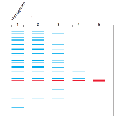
Biochemical purification of a protein from a cell extract often requires several purification steps involving various techniques. The purification process can be followed by gel electrophoresis of the starting protein mixture i.e. the cell homogenate and the fractions obtained from each subsequent purification step. Shown here are the schematic depictions of separation of proteins on a gel for the starting mixture of proteins (lane 1) and samples taken after each of the several purification steps.
If lanes 1 and 2 indicate the separation of proteins from the crude cell homogenate and after salt fractionation respectively, then which of the techniques are represented in lanes 3, 4 and 5 respectively?A . Ion exchange chromatography; affinity chromatography and gel filtration chromatographyB . Gel filtration chromatography; ion exchange chromatography and affinity chromatographyC . Affinity chromatography; ion exchange chromatography and gel filtration chromatographyD . Ion exchange chromatography; gel filtration chromatography and affinity chromatography
A student was performing enzyme purification in the laboratory for the first time. She began with 1200 mg of crude protein extract containing an enzyme of interest. At the beginning as well as at every purification step, she determined: i. Total protein content ii. Total units of enzyme present (also known as Activity) iii. Specific activity of enzyme (Units/mg protein).
A. In a very efficient purification process, which of the following is expected to happen? Choose the correct option I. i to increase, ii to remain constant, iii to increase. II. i and ii to increase, iii to remain constant. III. i to decrease, ii and iii to increase. IV. i, ii and iii to increase
B. Data (protein content and activity) obtained by her during the process is given in the table. Complete the table by filling in the specific activity, % yield of activity and purification factor values.
| Step | Protein (mg) | Activity (U) | Specific activity (U/mg) | % Yield of activity | Purification factor |
|---|---|---|---|---|---|
| 0 | 1200 | 800 | 0.67 | - | - |
| 1 | 600 | 600 | 1.0 | ||
| 2 | 200 | 180 | 0.9 | ||
| 3 | 30 | 150 | 5.0 | ||
| 4 | 20 | 148 | 7.4 |
C. Which of the above is the least effective purification step?
D. Which of the above is the most effective purification step?
E. Which step is indicative that enzyme of interest is partially inactivated?
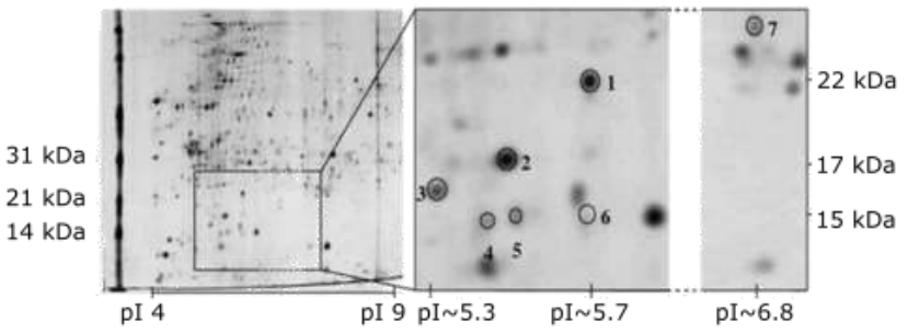
Plasmodium is a parasitic protozoan that is prevalent in tropical countries. Once the parasites invade host red blood cells, they multiply within 24 h. Fe2+ in red blood cell can react with free O2 as well as H2O2, causing formation of free radicals that can damage parasite cells. Unlike host cells, the parasites lack several enzymes of antioxidant defence systems. Scientists analysed the proteins from the parasite cytosol using two-dimensional gel electrophoresis (see image below). Then, using mass spectrometry and peptide mass fingerprinting, they identified 6 proteins (spots 1 to 6) corresponding to human peroxiredoxin, while one protein (spot 7) corresponding to Plasmodium peroxiredoxin.
Indicate if each of the following statements is True or False: A. Based on the presented data, Plasmodium peroxiredoxin is a multimeric protein. B. All human peroxiredoxin proteins have a positive net charge at physiological pH. C. Gel filtration chromatography is suitable for separation and purification of the six human peroxiredoxin proteins. D. Immunoaffinity chromatography can be used for separation of Plasmodium peroxiredoxin from other Plasmodium cytosolic proteins.
A professor wants to test his students’ knowledge on properties of amino acids. So he prepares a solution containing 0.9210g of unknown amino acid Z dissolved in 80mL deionized water and HCl equivalent to 3.2mL of 0.3024M NaOH solution. He now asks his students to take 25mL of the unknown amino acid solution in a 100ml beaker and measure the pH with a pH probe after adding 1mL of 0.3024M NaOH till 25mL NaOH has been added. The students make the following table:
| Volume of NaOH added (mL) | pH |
|---|---|
| 0.00 | 1.3 |
| 1.00 | 1.4 |
| 2.00 | 1.4 |
| 3.00 | 1.5 |
| 4.00 | 1.6 |
| 5.00 | 1.7 |
| 6.00 | 1.8 |
| 7.00 | 2.0 |
| 8.00 | 2.1 |
| 9.00 | 2.3 |
| 10.00 | 2.6 |
| 11.00 | 3.0 |
| 12.00 | 9.1 |
| 13.00 | 9.8 |
| 14.00 | 10.1 |
| 15.00 | 10.3 |
| 16.00 | 10.5 |
| 17.00 | 10.8 |
| 18.00 | 11.0 |
| 19.00 | 11.2 |
| 20.00 | 11.5 |
| 21.00 | 11.7 |
| 22.00 | 11.9 |
| 23.00 | 12.0 |
| 24.00 | 12.1 |
| 25.00 | 12.2 |
With this data, answer the following questions:
A. Plot graphs of pH vs Volume of NaOH added and find out value of pKa1, pKa2 and pI of amino acid Z.
B. Determine molecular weight of amino acid Z.
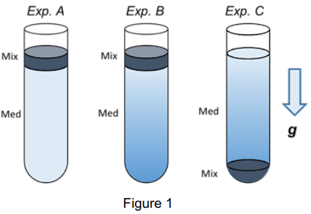
Here is a mixture containing viruses, globular proteins, and cell nuclei, which are all assumed to have similar densities of approximately 1.3 g/mL. We would like to separate them by using three different centrifugation methods as shown in Figure 1. The first method entails centrifugation of the mixture (Mix) after placing it on the top of a medium (Med) that has a uniform density (Exp. A). The second method (Exp. B) entails centrifugation of the mixture using a medium that has a density gradient ranging from 1.0 to 1.6 g/mL (from the top to the bottom). The final method entails the use of a centrifuge tube with the same density gradient as that in Exp. B, but the mixture is placed at the bottom of the tube (Exp. C). g indicates the direction of centrifugal forces given to the specimens.
A. In Exp. A, how are viruses, globular proteins, and nuclei supposed to sediment? Choose the most appropriate diagram from I, II, III, IV in Figure 2 that shows the sedimentation time courses of specimens
B. In Exp. A, choose appropriate lines from a, b, or c in the selected diagram that indicate the time courses of sedimentation of viruses, globular proteins and nuclei.
C. In Exp. B, how are viruses, globular proteins, and nuclei supposed to sediment? Choose the most appropriate diagram from I, II, III, IV in Figure 2 that shows the sedimentation time courses of the specimens.
D. In Exp. B, choose appropriate lines from a, b, or c in the selected diagram that indicate the time courses of sedimentation of viruses, globular proteins and nuclei.
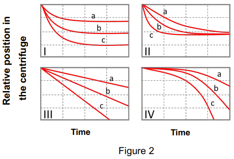
E. In Exp. C, how are viruses, globular proteins, and nuclei supposed to float? Choose the most appropriate diagram from I, II, III, IV in Figure 3 that shows the sedimentation time courses of the specimens.
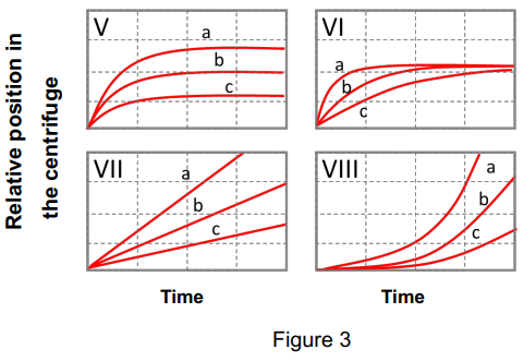
F. In Exp. C, choose appropriate lines from a, b, or c in the selected diagram that indicate the time courses of floating of viruses, globular proteins and nuclei.
Practice Problems
1. Membrane protein extraction. A researcher wants to extract an integral membrane protein while preserving its native folded structure for a functional assay. Which of the following is the most appropriate first choice: 1% SDS, 8 M urea, 1% Triton X-100 (a non-ionic detergent), or high salt concentration (2 M NaCl)?
Show answer
1% Triton X-100. Non-ionic detergents disrupt the lipid bilayer by inserting into it, releasing integral membrane proteins without denaturing them. SDS and urea are both strong denaturants (ionic and chaotropic, respectively) that would destroy native structure. High salt disrupts only ionic interactions and would not solubilise an integral (lipid-embedded) membrane protein at all; it releases peripheral membrane proteins instead.
2. A protein sample gives an A₂₆₀/A₂₈₀ ratio of 1.3, well below the ~1.8 expected for pure DNA. Propose the most likely explanation, and describe one purification step that would improve this ratio.
3. You need to purify a native (fully folded, active) tetrameric enzyme away from a mixture also containing free monomeric subunits of the same protein and several unrelated contaminant proteins of similar size. Which single technique from this page separates the intact tetramer from its own free monomers most directly, and why would SDS-PAGE alone fail to answer this specific question?