Q8 - Identifying DNA-Metabolism Enzymes from a Gel Assay
An in vitro experiment with enzymes involved in DNA metabolism was performed using the double-stranded DNA substrate shown in Figure 1. The upper strand of the DNA substrate is 50 bases, and the 5’ is labeled with blue fluorescent dye, while the lower strand is 100 bases, and the 3’ is labeled with red fluorescent dye. Fluorophore-labeled ends are chemically inert.
The reaction scheme is shown in Table 1. After each reaction, DNAs were separated using denaturing polyacrylamide gel electrophoresis to separate denatured single-stranded DNAs (Figure 2, the purple color indicates the presence of both DNA strands at the same position).
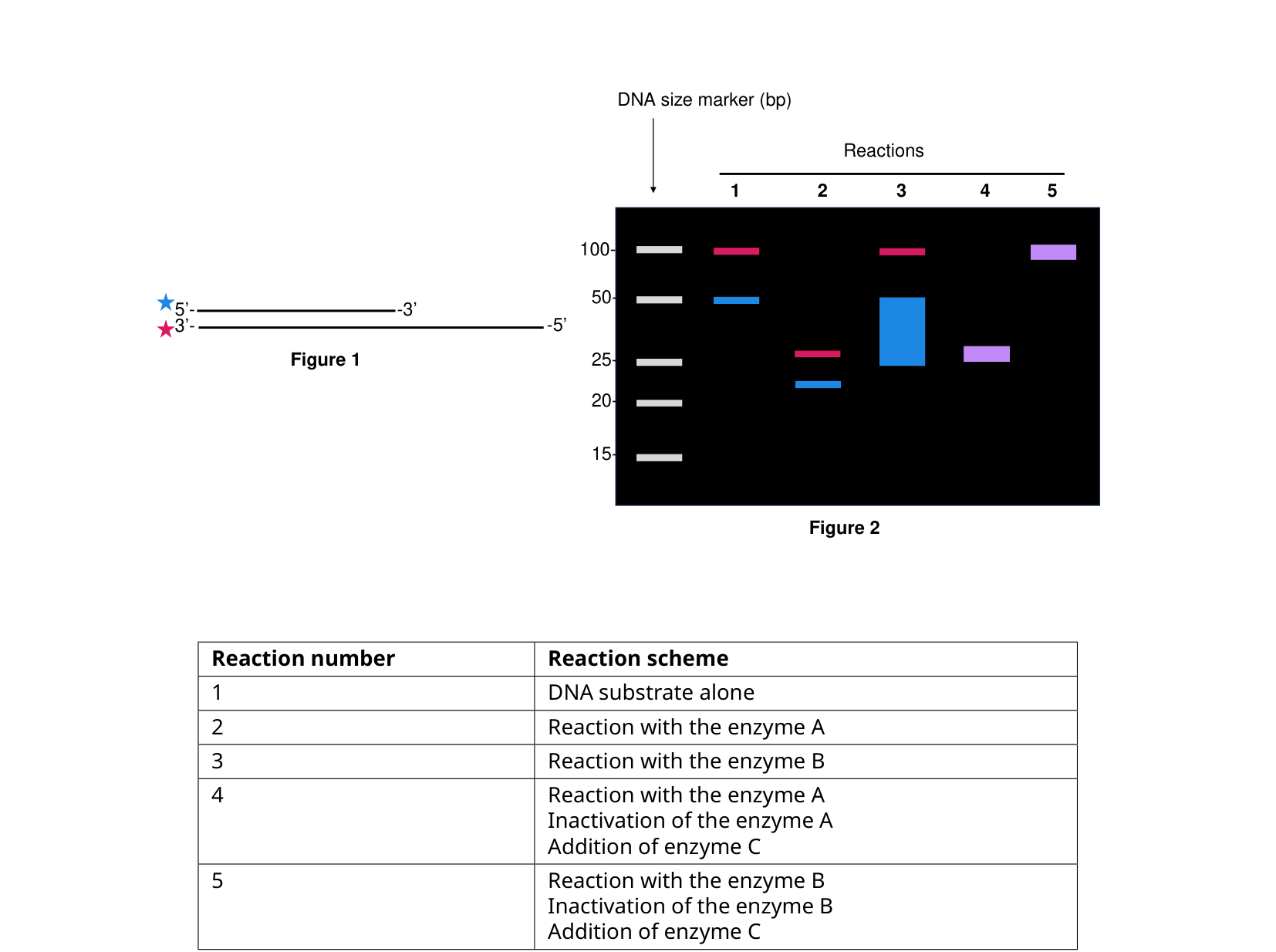 Figure 1 (substrate diagram) and Figure 2 (gel result), with Table 1 (reaction scheme): reaction 1 = DNA substrate alone; reaction 2 = + enzyme A; reaction 3 = + enzyme B; reaction 4 = + enzyme A, then inactivate A, then + enzyme C; reaction 5 = + enzyme B, then inactivate B, then + enzyme C.
On your answer sheet, indicate “T” for true statements and “F” for false ones.
Question reproduced from IBO 2024, Theoretical Exam Part B, licensed under CC BY-NC-SA 4.0 - attributed to the International Biology Olympiad. Open the full exam PDF