Q10 - 3D Bioprinting - Cell Viability, Bioluminescence, and Confocal Imaging
To create in vitro analogues of live tissue, 3D bioprinters use what is called bioink. Bioink formulations combine live cells with different kinds of biocompatible materials that can be polymerized after extrusion from a printing needle, letting cells proliferate and form 3D structures.
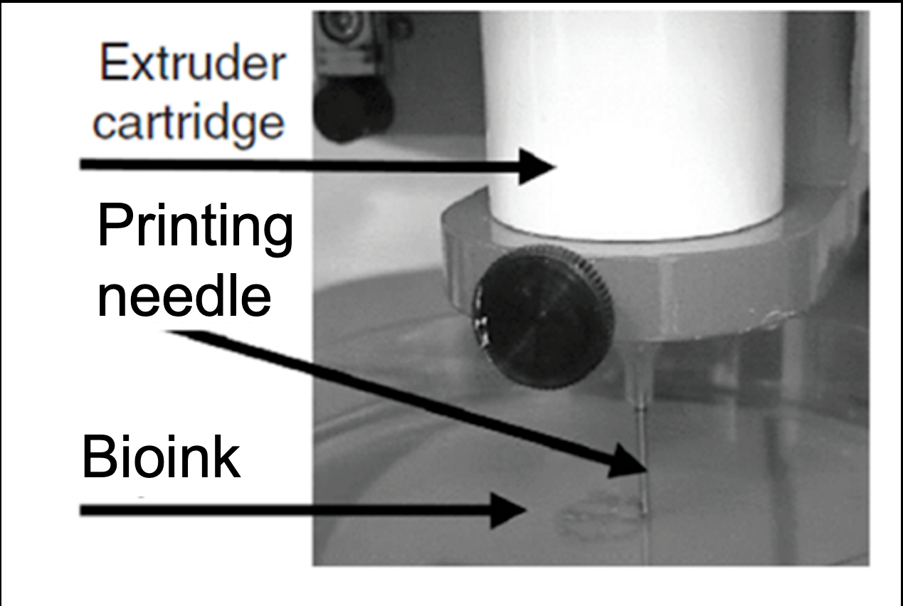 Figure 1. The 3D printer of living tissues - extruder cartridge, printing needle, and bioink being deposited.
The viability of cells post-printing can be evaluated by daily monitoring of bioluminescence, which essentially measures intracellular ATP. Due to shear stress associated with the extrusion process, cell viability and proliferation within printed constructs can be adversely affected by needle size and extrusion pressure. Bioluminescence data and confocal images were acquired from constructs printed with bioink containing either fibroblasts or myocytes; after several days of culturing, both construct types were stained with the same markers.
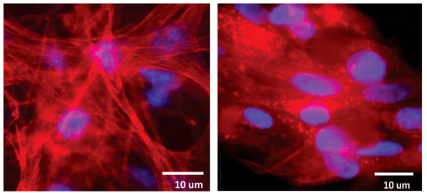 Figure 2. Confocal images from tissue constructs printed with bioink containing fibroblasts (left) or myocytes (right) - red cytoskeletal staining, blue nuclear staining.
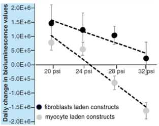 Figure 3. Daily change in bioluminescence values vs. pressure (psi). Both fibroblast- and myocyte-laden constructs decline as pressure increases, with myocyte-laden constructs declining faster, dropping to negative values at higher pressures.
Using the information and figures, determine whether the following statements are true or false:
Question reproduced from IBO 2022, Theoretical Paper 1, licensed under CC BY-NC-SA 4.0 - attributed to the International Biology Olympiad. Open the full exam PDF · Community solutions (unofficial)