Q9 - Organ-on-a-Chip - Lung-on-a-Chip & Pulmonary Oedema Model
Tissue engineering is trying to develop miniature blocks of tissue from multiple cell types using an approach called ‘organ-on-a-chip’. This technology enables in vitro testing of patient-specific effects of different drugs or treatments. Cells collected from a patient are first turned into induced pluripotent stem cells (iPSCs), then differentiated and seeded into specially designed chambers.
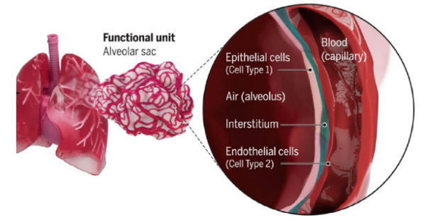 Figure 1. The endothelial barrier of alveoli - epithelial cells facing air (alveolus), separated from endothelial cells facing blood (capillary) by a thin interstitium.
This ’lung-on-a-chip’ models pulmonary oedema progression. Endothelial barrier function was quantified by measuring alveolar-capillary permeability to a fluorescently labeled inulin, a large polysaccharide molecule. Inulin was introduced into the lower chamber together with interleukin-2 (IL-2), one of the major cytokines.
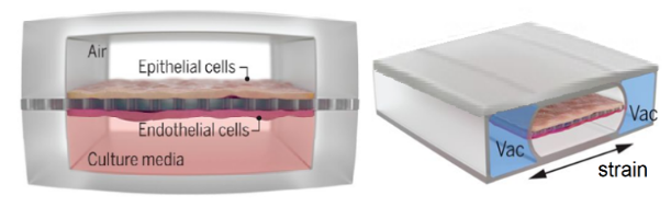 Figure 2. The chip chamber used in the ‘organ-on-a-chip’ device: epithelial cells over endothelial cells, separated by a porous membrane, with vacuum channels applying cyclic mechanical strain.
Images next to the graph below illustrate immunostaining patterns for junctional proteins - epithelial occludin (green) and endothelial VE-cadherin (red) - in control conditions and after exposure to cyclic mechanical strain (10% at 0.2 Hz) along with IL-2 for 3 days. White arrows indicate intercellular gaps.
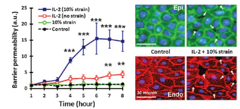 Figure 3. Barrier permeability (a.u.) vs. time (hours) for four conditions: IL-2 + 10% strain (highest), IL-2 no strain, 10% strain alone, and control (both near baseline). Right: immunostaining for occludin (Epi, green) and VE-cadherin (Endo, red) - control vs. IL-2 + strain, with white arrows marking intercellular gaps.
Based on the data provided, indicate whether the following statements are true or false:
Question reproduced from IBO 2022, Theoretical Paper 1, licensed under CC BY-NC-SA 4.0 - attributed to the International Biology Olympiad. Open the full exam PDF · Community solutions (unofficial)