Cell Junctions, Extracellular Matrix & Cell Death
Overview
This closing Cell Biology page covers how cells physically connect to each other and to their surroundings, and how cells die, either as a controlled, regulated program or as uncontrolled damage. Both topics share a theme running through this whole tier: the cell actively manages its boundaries and its own survival, rather than these being passive default states.
Key Concepts
Cell junctions: three types, three distinct jobs
Animal cells form three structurally and functionally distinct junction types, each solving a different problem:
- Tight junctions: seal adjacent cells together via interlocking transmembrane proteins (claudins, occludins), creating a nearly impermeable barrier that blocks even small molecules from diffusing between cells (the paracellular route). Found in epithelia that must maintain a strict compartmental separation, e.g. intestinal epithelium separating gut lumen from the bloodstream, or the blood-brain barrier, forcing all transport across the epithelial sheet through the cells themselves (transcellular, regulatable) rather than around them.
- Anchoring junctions (adherens junctions and desmosomes): mechanically link the cytoskeletons of adjacent cells (via cadherin proteins spanning the intercellular space) or link a cell to the extracellular matrix (via integrins, in hemidesmosomes and focal adhesions). Adherens junctions couple to actin filaments; desmosomes couple to intermediate filaments (see Cytoskeleton & Motor Proteins), this distinction matters functionally, since intermediate-filament-anchored desmosomes provide the greater tensile/mechanical resilience needed in tissues under high mechanical stress (e.g. skin, cardiac muscle).
- Gap junctions: form direct cytoplasmic channels (built from connexin proteins arranged into a connexon) between adjacent cells, allowing small molecules and ions to pass directly cell-to-cell without crossing either plasma membrane individually. This provides direct electrical/chemical coupling: critical in cardiac muscle for coordinated, synchronised contraction, since an action potential can propagate directly cell-to-cell through gap junctions rather than requiring a slower cell-surface-receptor-mediated relay each time.

Extracellular matrix (ECM)
The ECM is the network of secreted extracellular proteins and polysaccharides surrounding animal cells, providing structural support, anchorage points, and a substrate that itself carries signaling information (transduced into the cell via integrin receptors, linking the ECM back to intracellular signaling, see Cell Signaling & Communication). Major components: collagen (the most abundant ECM protein, providing tensile strength via its triple-helix structure; see Protein Structure, Folding & Function), proteoglycans (core proteins heavily decorated with glycosaminoglycan chains, whose dense negative charge draws water in, resisting compression; see Carbohydrate Chemistry & Biology), and fibronectin (links ECM components to cell-surface integrins, coordinating cell adhesion, migration, and shape).
Apoptosis: programmed cell death
Apoptosis is a tightly regulated, energy-requiring (ATP-dependent) form of programmed cell death, essential for normal development (e.g. sculpting digits by removing interdigital webbing, eliminating self-reactive immune cells) and ongoing tissue homeostasis (removing damaged or superfluous cells without triggering inflammation). Two convergent activation pathways:
- Intrinsic (mitochondrial) pathway: triggered by internal stress signals (DNA damage, growth factor withdrawal), leading to mitochondrial outer membrane permeabilisation and release of cytochrome c into the cytoplasm, a striking example of a normally strictly compartmentalised mitochondrial protein (see Mitochondria & Chloroplasts) being repurposed as a death signal once released.
- Extrinsic (death receptor) pathway: triggered by extracellular ligand binding to a cell-surface “death receptor” (e.g. Fas), directly activating the downstream protease cascade without a mitochondrial step.

Both pathways converge on activating a proteolytic cascade of caspases, cysteine proteases that cleave specific target proteins to systematically dismantle the cell: the cytoskeleton is disassembled, chromatin is condensed and DNA is fragmented, and the cell breaks into membrane-bound apoptotic bodies. Externalised phosphatidylserine on the outer membrane leaflet (see Lipids & Membrane Biochemistry for the lipid-asymmetry chemistry) serves as an “eat me” signal, allowing clean phagocytic clearance of apoptotic bodies without releasing cellular contents into the surrounding tissue: the critical distinction that keeps apoptosis non-inflammatory.
Necrosis: uncontrolled cell death
Necrosis results from acute, overwhelming cellular injury (e.g. ischemia, physical trauma, toxins) rather than a regulated program. The cell swells (osmotic/ionic regulation fails), the plasma membrane ruptures, and cellular contents (including lysosomal enzymes and pro-inflammatory intracellular molecules) spill into the surrounding tissue, triggering inflammation, the direct opposite of apoptosis’s clean, non-inflammatory clearance. This inflammation vs. no-inflammation distinction is the single most important functional/exam contrast between the two death modes.

Autophagy: self-digestion, not necessarily death
Autophagy is a distinct process from both of the above: a double-membrane autophagosome forms around damaged organelles, protein aggregates, or (under starvation) bulk cytoplasmic material, then fuses with a lysosome (see Endomembrane System & Protein Trafficking) for degradation and recycling of the components. Autophagy is primarily a survival mechanism, allowing a starving cell to recycle its own components for energy, or removing damaged organelles/aggregated proteins before they cause harm, though sustained or excessive autophagy can also contribute to cell death in some contexts, which is why it is sometimes (imprecisely) grouped alongside apoptosis and necrosis as a “third type of cell death” despite its primary role being protective rather than destructive.
Comparative Structures
| Feature | Apoptosis | Necrosis | Autophagy |
|---|---|---|---|
| Regulated/programmed? | Yes | No (acute injury response) | Yes |
| Energy requirement | ATP-dependent | Not dependent (often occurs as ATP fails) | ATP-dependent |
| Membrane integrity | Preserved until clean packaging into apoptotic bodies | Ruptures, contents spill | Preserved (self-contained autophagosome) |
| Inflammatory? | No | Yes | No |
| Primary trigger | Internal stress signal or death receptor | Acute overwhelming injury | Nutrient stress, damaged organelle/protein clearance |
| Key molecular players | Caspases, cytochrome c | (Passive failure, no dedicated pathway) | Autophagosome, lysosome fusion |
Common Exam Questions
- “Why is apoptosis non-inflammatory but necrosis is?”: the key mechanistic distinction is whether cellular contents are released into surrounding tissue (necrosis) or cleanly packaged and phagocytosed before any leakage occurs (apoptosis), this is the testable causal chain, not simply “apoptosis is controlled.”
- “What does externalised phosphatidylserine signal, and why is its normal location relevant?”, tests the connection back to membrane lipid asymmetry (normally maintained by flippases) as the mechanistic basis of the “eat me” signal.
- Distinguishing intrinsic vs. extrinsic apoptotic pathway triggers (internal stress/mitochondrial vs. external death-receptor ligand) is a frequent classification question.
- “Which junction type would be disrupted by a mutation in connexin?”: gap junctions specifically; questions naming a specific protein (cadherin, claudin, connexin, integrin) test direct recall of which junction type that protein belongs to.
Visual Reference
Interactive
- A cell-death pathway decision tree: given a scenario description (e.g. “acute physical trauma” vs. “developmental digit sculpting” vs. “nutrient starvation”), select which death/survival mode applies and see the correct molecular pathway highlighted.
Static
(Static images are placed inline in Key Concepts above, next to the concept each one illustrates, rather than collected here.)
Practice Challenge
Competition-sourced practice questions for this topic, graded by difficulty. Click the Solution tab to reveal each answer.
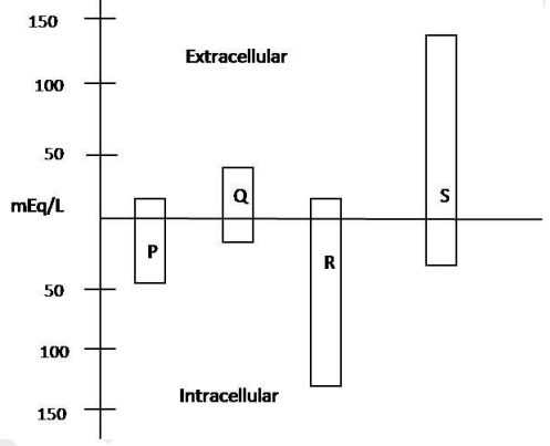
Concentrations of cations & anions present in intracellular & extracellular fluids of human body are depicted in the graph.
P, Q, R and S respectively are:A. K+, Na+, Cl- and organic anions B. Proteins, bicarbonates, K+ and Na+C. Mg++, proteins, bicarbonates and Na+D. Ca++, lactate, K+ and Cl-
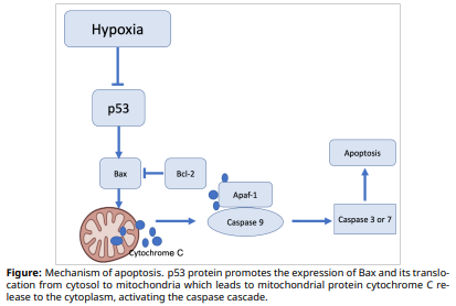
Hypoxia is a hallmark of solid tumors and plays a critical role in different steps of tumor progression. To develop an effective treatment against malignant tumors, it is important to understand how cancer cells that develop under hypoxic conditions, avoid apoptosis. The figure shows the mechanism of apoptosis.
Using the information and figure, determine whether the following statement are true or false:A. Hypoxia in cancer cells suppresses apoptosis.
B. Upon Bcl-2 overexpression, the integrity of the mitochondrial membranes is preserved, which in return prevents apoptosis.
C. It is necessary to suppress p53 to ensure the apoptosis of cancer cells.
D. Deficiency of Bax protein promotes apoptosis.
Sir Alastair Currie (1921-1994), Sir John Sulston (1942-present) and colleagues discovered how cells can commit controlled suicide (apoptosis). Apoptosis is executed by Caspase enzymes. Cyclohexamide (CH), which inhibits ribosomes, was used to induce apoptosis in cells treated with the hormone bone morphogenetic protein-9 (BMP9). To measure the number of apoptotic cells after treatment, inactive luciferase substrate, and the light-emitting enzyme luciferase, were added.
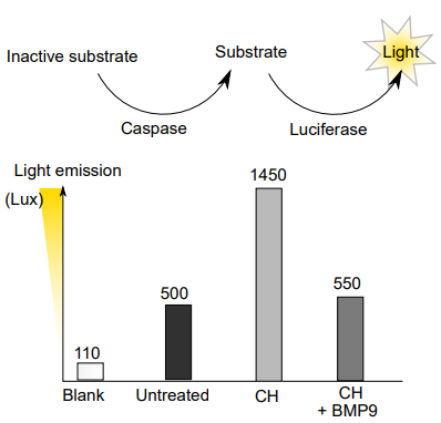
(I) Find reduction (in %) in the number of apoptotic cells when treated with CH + BMP9 compared to only CH treatment
The effect of knocking out specific genes in worms on apoptosis (programmed cell death) was studied.
| Gene | Manipulation | Annexin-V labelled cells |
|---|---|---|
| ced9 | knock-out | Increased |
| ced4 | knock-out | Reduced |
| ced3 | knock-out | Reduced |
| egl1 | knock-out | Reduced |
| ced4 & ced9 | knock-out | Reduced |
| ced9 & ced3 | knock-out | Reduced |
| ced9 & egl1 | knock-out | Increased |
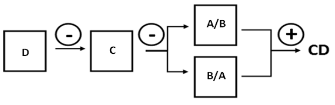
Write the letters A-D corresponding to the genes into the correct rectangle in the model of the control of cell death (CD) along with whether its interaction is activating (+) or repressing (-) in the circles on the arrows. A. Ced3B. Ced4C. Ced9D. Egl1
Practice Problems
1. A tissue sample shows widespread cell swelling, membrane rupture, and a strong local inflammatory response following an ischemic injury. Which cell death mode is most consistent with these findings, and what specific observation would most clearly rule out the alternative?
Show answer
Necrosis. The combination of membrane rupture, cell swelling, and inflammation is the necrosis signature; apoptosis would instead show intact membrane-bound apoptotic bodies with no significant inflammatory response, since apoptotic content is cleanly phagocytosed before any leakage. The clearest distinguishing observation would be whether cellular contents (e.g. lysosomal enzymes, intracellular proteins) are found extracellularly: present in necrosis, absent in properly executed apoptosis.
2. A cardiac tissue sample shows a mutation eliminating functional connexin protein. Predict the physiological consequence for coordinated heart muscle contraction, and explain the mechanism.
3. Explain why autophagy is sometimes described as “a cell eating itself to survive” rather than simply classified as a form of cell death, referencing its typical trigger and outcome.