Q71 - Hippo Pathway Signaling in Blastocyst Implantation
One week after fertilization, human embryos implant into the uterus. In order to implant, the embryo needs to develop into a blastocyst (blastoid), a spherical structure covered with cells. Researchers simulated a blastocyst model, using pluripotent stem cells. They also modeled the endometrial layer of the uterus in the dish and blastocyst implantation in the endometrium (Figure 1). They hormonally stimulated endometrial cells, and tested blastocyst attachment (Figure 2). In order to check the effect of the Hippo pathway on blastoid development, researchers tested them under exposure to different concentrations of lysophosphatidic acid (LPA) (a Hippo-pathway inhibitor) (Figure 3). YAP1 is the downstream effector that activates cavitation, after Hippo inhibition, by binding to the TEAD transcription factor. The researchers tested different settings on the appearance of cavitation, an important event in blastocyst formation. They used two different YAP1-overexpressing (OE) blastocysts, overexpressing two different YAP1-mutants: one has a mutation in the TEAD binding site (S94A), while the other is constitutively active (5SA) (Figure 3).
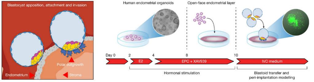 Figure 1. Diagram of attachment of the blastoid to endometrial cells (left) and the method for producing the endometrial organoids used for in vitro implantation modeling (right).
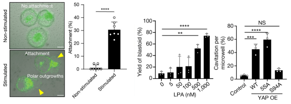 Figure 2 (left): blastoid attachment to non-stimulated vs. hormonally stimulated endometrial cells. Figure 3 (right): blastoid yield as a function of LPA (Hippo-pathway inhibitor) concentration, and cavitation per microwell for control cells versus cells overexpressing wild-type, 5SA, or S94A YAP1.
Use the information and figures 1-3 to determine whether the following statements are true or false.
Question reproduced from IBO 2022, Theoretical Paper 2, licensed under CC BY-NC-SA 4.0 - attributed to the International Biology Olympiad. Open the full exam PDF · Community solutions (unofficial)