Cell Signaling & Communication
Overview
Cells constantly receive and interpret chemical signals from their environment and from other cells, converting a single molecular binding event at the cell surface into a coordinated intracellular response, often affecting thousands of downstream molecules from one bound signal. This page covers the three-stage framework (reception, transduction, response) that organises all cell signaling, then the two dominant surface receptor classes and their downstream cascades.
Key Concepts
The three-stage framework
Every signaling pathway, regardless of specific molecular details, follows the same logical sequence:
- Reception: a signaling molecule (ligand) binds a specific receptor, either at the cell surface (for large or hydrophilic ligands unable to cross the plasma membrane) or intracellularly (for small hydrophobic ligands, e.g. steroid hormones, that diffuse directly across the membrane).
- Transduction: ligand binding triggers a series of molecular changes, often a relay of sequential protein modifications, that carries and typically amplifies the signal from the receptor into the cell interior.
- Response: the transduced signal ultimately alters cell behaviour: activating/inhibiting an enzyme, opening a channel, or (very commonly) altering gene transcription via activation of a transcription factor (see Gene Regulation: Eukaryotic & Epigenetics).
G-protein-coupled receptors (GPCRs)
GPCRs are the largest receptor family, defined by seven transmembrane α-helices and an associated intracellular heterotrimeric G-protein (Gα, Gβ, Gγ subunits). In the resting state, Gα binds GDP; ligand binding to the receptor causes a conformational change that acts as a guanine nucleotide exchange factor (GEF), prompting Gα to release GDP and bind GTP instead. GTP-bound Gα then dissociates from Gβγ, and each can independently activate downstream effectors. Gα has intrinsic GTPase activity that eventually hydrolyses its own bound GTP back to GDP, terminating the signal and allowing reassembly of the inactive heterotrimer, a built-in, self-limiting timer on signal duration.

A common downstream effector is adenylyl cyclase, activated by one class of Gα (Gαs) to convert ATP into the second messenger cyclic AMP (cAMP), which in turn activates protein kinase A (PKA), phosphorylating a range of downstream target proteins. A different Gα class (Gαi) inhibits adenylyl cyclase instead: the same receptor family can therefore either raise or lower cAMP levels depending on which G-protein subtype it’s coupled to, a key source of signaling specificity across the many distinct GPCRs a single cell may express.
Receptor tyrosine kinases (RTKs)
RTKs are single-pass transmembrane receptors with an intrinsic tyrosine kinase enzymatic domain on their cytoplasmic side. Ligand binding (often a growth factor) causes receptor dimerisation, bringing two cytoplasmic kinase domains into proximity so each phosphorylates tyrosine residues on the other: trans-autophosphorylation. These phosphotyrosines then serve as docking sites for intracellular signaling proteins carrying SH2 domains, which recognise phosphotyrosine specifically, nucleating a multi-protein signaling complex at the activated receptor. RTK signaling is central to cell growth and proliferation control (e.g. the Ras/MAPK cascade, a downstream RTK pathway with major relevance to cancer biology, since constitutively active Ras mutants are among the most common oncogenic mutations found in human tumours).

Second messengers: amplifying and diversifying the signal
Small, rapidly diffusible intracellular molecules that relay and amplify a signal beyond the initial receptor-ligand binding event:
- cAMP: produced by adenylyl cyclase (above), activates PKA.
- Ca²⁺: normally held at very low cytoplasmic concentration by active pumping into the ER/sarcoplasmic reticulum and extracellularly; signal-triggered release (e.g. via IP₃ opening ER Ca²⁺ channels, below) produces a sharp, fast concentration spike detectable by Ca²⁺-binding proteins like calmodulin.
- IP₃ and DAG: generated together by phospholipase C (PLC) cleaving the membrane phosphoinositide PIP₂ (see Lipids & Membrane Biochemistry for the lipid chemistry): IP₃ diffuses into the cytoplasm and triggers ER Ca²⁺ release; DAG stays membrane-associated and activates protein kinase C (PKC). One receptor-binding event thus produces two parallel second-messenger branches from a single enzymatic cleavage step.

The core reason second messengers matter functionally, beyond relaying the signal at all, is amplification: one activated receptor can activate many G-protein/enzyme molecules, each of which generates many second-messenger molecules, each of which can activate many kinase molecules: a small number of surface-binding events can therefore produce a very large, fast intracellular response, and this multiplicative cascade structure is the general mechanistic reason cells can respond so quickly and strongly to low concentrations of an extracellular signal.
Signal termination
Every activating step described above has a matched inactivating mechanism, Gα’s intrinsic GTPase activity, phosphatases that remove activating phosphates added by kinases, and active Ca²⁺ resequestration into the ER, because a signaling pathway that only turns on and never off cannot function as a regulated switch. Dysregulation of termination steps (e.g. a Ras mutant with impaired GTPase activity, staying locked in the “on,” GTP-bound state) is a recurring theme in disease, not just activation-side mutations.
Comparative Structures
| Feature | GPCR pathway | RTK pathway |
|---|---|---|
| Receptor structure | 7-transmembrane helix | Single-pass transmembrane, intrinsic kinase domain |
| Activation mechanism | G-protein GEF activity, GDP→GTP exchange | Ligand-induced dimerisation, trans-autophosphorylation |
| Immediate downstream event | Second messenger production (e.g. cAMP, IP₃/DAG) | SH2-domain protein docking at phosphotyrosines |
| Built-in “off switch” | Gα intrinsic GTPase activity | Phosphatase removal of phosphotyrosines |
| Classic downstream pathway | cAMP/PKA, or PLC/IP₃-DAG | Ras/MAPK |
Common Exam Questions
- “Explain how a single ligand-binding event produces a large, fast cellular response”: the expected answer is the multiplicative amplification through the receptor → G-protein/second-messenger → kinase cascade, not simply “the signal travels into the cell.”
- “Why does a Ras mutant with impaired GTPase activity act as an oncogene?”: tests understanding that GTP hydrolysis is the off switch; losing it locks the pathway in a constitutively active state, mimicking permanent growth-factor stimulation.
- Distinguishing GPCR from RTK signaling by the type of receptor structure and the nature of the immediate post-binding event (G-protein activation vs. receptor dimerisation/autophosphorylation) is a frequent classification question.
Visual Reference
Interactive
- A signal amplification cascade animator: click “ligand binds,” then watch each downstream stage (receptor → G-protein/second messenger → kinase → target proteins) activate in sequence, with a running count of “molecules activated” at each stage to make the amplification concept quantitatively visible.
Static
(Static images are placed inline in Key Concepts above, next to the concept each one illustrates, rather than collected here.)
Practice Challenge
Competition-sourced practice questions for this topic, graded by difficulty. Click the Solution tab to reveal each answer.
Due to dynamic phosphorylation-dephosphorylation process, almost all signalling pathways are reversible and in many cases phosphorylation activates the signalling. Which combination of following conditions will inhibit such a signalling pathway in operation? i. Application of kinase inhibitor.ii. Treatment with a phosphatase inhibitor.iii. Phosphorylated amino acid residue binder recruiting a phosphatase.iv. Binding of a phosphorylated amino acid residue recognizing kinase.v. An interaction stabilizing the half-life of the phosphorylated amino acid residue.
A. Only i and iiiB. Only ii, iv, and vC. Only i, iii, and ivD. Only ii, iii, and v
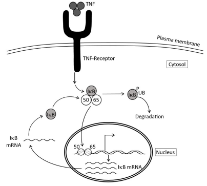
Signal transduction events through transmembrane receptors in cells often culminate in the activation of a transcription factor. One such example is the activation of NF-κB (Nuclear Factor Kappa B) by TNF-α (Tumor Necrosis Factor-alpha). NF-κB is a heterodimer of two subunits, p50 and p65, and it is retained in the cytoplasm by its inhibitor IκB-α. TNF signaling phosphorylates IκB-α leading to its degradation by ubiquitination. As a result, free NF-κB and its translocation into the nucleus, where it binds to a responsive promoter and activates gene expression. Activation of NF-κB by the TNF is transient in nature because one of the genes that NF-κB activates is the gene for IκB-α itself. In one experiment, activation of NF-κB by TNF was studied in a macrophage cell line. From the above experiment, cytoplasmic and nuclear extracts were prepared, proteins were separated on SDS-PAGE and Western blotting was performed using antibodies against p65 and IκB-α proteins. Also, antibodies against α-actin and lamin were used to demonstrate the purity of the cytoplasmic and nuclear fractions as well as loading control.
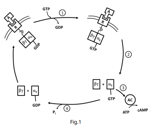
G protein-coupled receptors (GPCRs) interact with G proteins that subsequently affect cell function through the generation of second messengers. Cyclic AMP (cAMP) generated by adenylyl cyclase controls cell functions via activation of protein kinases. GPCRs may either activate or inhibit the cyclase through the G proteins Gs and Gi, respectively. The difference between Gs and Gi resides in the 𝛼 subunit, which binds and hydrolyses GTP. The Gs protein cycle is illustrated below (Fig. 1).
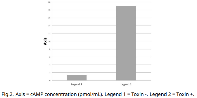
A lab is working on a pair of newly identified GPCRs, “GPCR-A” and “GPCR-B”. Each binds the same small ligand with the same affinity but activates different G-proteins that act on adenylyl cyclase. When activated, GPCR-A causes an increase in adenylyl cyclase activity, while GPCR-B causes a decrease in adenylyl cyclase activity. They have a cell line that expresses both GPCR-A, GPCR-B, the corresponding G-proteins, and adenylyl cyclase. There is a basal level of adenylyl cyclase activity that produces a baseline cAMP concentration. A member of the lab studying a pathogenic bacterium has discovered that it secretes a toxin that interferes with the mentioned signalling pathway. To determine how this toxin acts, she did an experiment in which she looked at intracellular cAMP levels in untreated and toxin treated cells (the original ligand of the receptors was not added in either of the experiments).
(I) Indicate whether each of the following mutations increase (+), not changed (0) or decrease (-) the intracellular levels of cAMP upon ligand addition in the absence of the toxin? Both GPCR-A and GPCR-B bind the same ligand.
A. A mutation in GPCR-A that prevents G protein activation.
B. A mutation in GPCR-B that prevents G protein activation.
C. A mutation in Gs that prevents release of bound GDP.
D. A mutation in Gi that prevents release of bound GDP.
E. A mutation in Gs that prevents GTP hydrolysis.F. A mutation in Gi that prevents GTP hydrolysis.
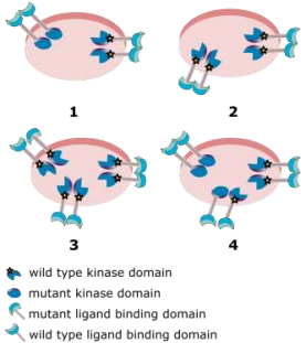
Two different mutant forms of a gene that encodes a cell surface receptor tyrosine kinase (RTK) are separately inserted into vectors. One mutant encodes a protein with a non-functional kinase domain, and the other lacks a functional ligand binding domain. Each vector is separately introduced into normal cells that can express wild type RTK from their endogenous genes.It is known that the cells used have a high capacity for RTK receptors on their surface, that ligands bind to monomeric forms of receptor proteins and that heterodimeric receptors are inactive in signalling. The diagrams below depict four cell types identified. Each diagram shows the only receptor forms observed on the respective cell types in the ratio that they were observed.The experiments were performed under non-saturating concentrations of ligand.
Indicate if each of the following statements is true or false:A. In type 1 cells, the mutant receptor with the non-functional kinase domain will interfere with signalling by the cells’ normal RTK.
B. In type 2 cells, the mutant RTK lacking functional ligand binding domain will be inactive for signalling, but will not interfere with normal signalling mediated by the cells’ own receptor tyrosine kinases.
C. Equal levels of signalling will be achieved by type 3 and type 4 cells.
D. The effects of mutant RTKs of cell type 2 and cell type 3 on levels of signalling by the cells’ own normal RTKs will be the same.
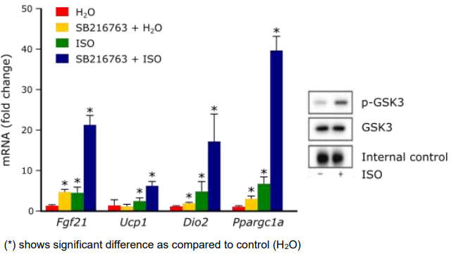
Brown adipose tissue (BAT) activation through β-adrenergic signalling pathway is associated with expression of thermogenic genes and the process of thermogenesis. Glycogen synthase kinase-3 (GSK3) acts as a regulator of β-adrenergic signalling in brown adipocytes. ISO is also a chemical stimulator of BAT activation; its effect on GSK3 is shown by the Western blot below (p-GSK3 is phosphorylated form of GSK3). SB216763 is an inhibitor of GSK3. Effects of these two agents on the expression of thermogenic genes (Fgf21, Ucp1, Dio2, and Ppargc1a) are shown in the Figure below
Indicate if each of the following statements is true or false:A. GSK3 acts as a negative regulator of β-adrenergic signalling in brown adipocytes.
B. Phosphorylation of GSK3 causes decreased expression of Fgf21.
C. SB216763 may prevent diet-induced obesity.
D. Use of SB216763 and ISO together cause much higher increase in the number of Ppargc1a mRNA transcripts as compared to Fgf21 mRNA transcripts.
Practice Problems
1. A cell is treated with cholera toxin, which locks Gαs in its GTP-bound (active) state by blocking its intrinsic GTPase activity. Predict the effect on intracellular cAMP levels, and explain the mechanism.
Show answer
Intracellular cAMP rises persistently and abnormally high. Locking Gαs in the GTP-bound state removes the pathway’s normal “off switch” (GTP hydrolysis back to GDP), so Gαs continues activating adenylyl cyclase indefinitely rather than for the normal, self-limited signal duration. cAMP accumulates continuously rather than transiently. (This is the actual mechanism of cholera toxin’s pathology in intestinal epithelial cells, driving massive fluid secretion.)
2. Two different GPCRs in the same cell both bind their respective ligands, but one raises intracellular cAMP while the other lowers it. Explain how this is possible given that both are structurally GPCRs.
3. An RTK-family growth factor receptor carries a mutation preventing dimerisation upon ligand binding, but the kinase domain itself is fully catalytically functional in isolation. Predict the effect on downstream Ras/MAPK signaling, and explain why an intact kinase domain alone is insufficient.