Cell Cycle, Mitosis & Meiosis
Overview
Scope note: this page covers the mechanics of how chromosomes move and divide: cell cycle regulation, mitotic stages, and the chromosome behaviour of meiosis I and II. The genetic consequences of meiosis, independent assortment (see Mendel’s Laws & Probability in Genetics), crossing-over-derived recombination frequency and linkage mapping (see Linkage, Recombination & Genetic Mapping), are covered in Genetics, not repeated here; this page is the mechanistic foundation those topics build on.
Key Concepts
The cell cycle and its checkpoints
The eukaryotic cell cycle divides into interphase (G1, S, G2: the cell grows and duplicates its genome) and M phase (mitosis and cytokinesis: the cell actually divides). G1 is the primary growth phase; S phase is when DNA replication occurs (see DNA Structure & Replication), producing sister chromatids; G2 is a second growth/preparation phase before division.
Progression is controlled by cyclin-dependent kinases (CDKs), whose activity requires binding a regulatory cyclin subunit: cyclin levels rise and fall across the cycle (via regulated synthesis and, critically, ubiquitin-mediated degradation, see Protein Structure, Folding & Function), while CDK protein levels stay roughly constant; it is this oscillating cyclin availability, not CDK abundance, that drives the cycle’s directionality. Three major checkpoints enforce quality control:
- G1/S checkpoint (“restriction point”): checks cell size, nutrient availability, and DNA integrity before committing to replicate the genome; the primary point at which a cell decides whether to proceed through the cycle at all, or exit into a non-dividing resting state (G0).
- G2/M checkpoint: verifies DNA replication completed correctly and checks for DNA damage before allowing entry into mitosis.
- Spindle assembly checkpoint (metaphase checkpoint): verifies every chromosome’s kinetochore is properly attached to spindle microtubules from both poles before allowing anaphase onset: a single unattached kinetochore is sufficient to halt the entire cell cycle, preventing catastrophic chromosome missegregation.
Loss of checkpoint function is a recurring cancer mechanism: tumour suppressor genes like p53 function largely as checkpoint enforcers (triggering cell-cycle arrest or apoptosis in response to DNA damage), so their loss allows damaged cells to continue dividing unchecked.

Mitosis: the stages
Mitosis divides a duplicated genome (sister chromatids) into two genetically identical daughter nuclei:
- Prophase: chromatin condenses into visible chromosomes (each still consisting of two sister chromatids joined at the centromere); the mitotic spindle begins forming as centrosomes migrate toward opposite poles.
- Prometaphase: the nuclear envelope breaks down (requiring depolymerisation of the nuclear lamina, see Cytoskeleton & Motor Proteins); spindle microtubules invade the nuclear region and begin attaching to kinetochores via dynamic instability-driven search-and-capture (see Cytoskeleton & Motor Proteins).
- Metaphase: all chromosomes align at the metaphase plate, each properly attached to microtubules from both spindle poles (bi-orientation), the spindle assembly checkpoint is satisfied at this point.
- Anaphase: sister chromatids separate simultaneously and are pulled toward opposite poles, driven by microtubule shortening at the kinetochore and by motor-protein-driven pole separation.
- Telophase: chromosomes decondense, nuclear envelopes reform around each set of chromosomes, and the spindle disassembles.
- Cytokinesis (overlapping with late mitosis, mechanistically separate from it): in animal cells, an actin-myosin contractile ring assembles at the cell cortex and constricts, pinching the cell in two (see Cytoskeleton & Motor Proteins for the actin/myosin mechanism); in plant cells, which have a rigid cell wall preventing this pinching mechanism, a cell plate instead forms from the inside outward via vesicle fusion, eventually forming a new cell wall.

Meiosis: chromosome mechanics
Meiosis produces four genetically distinct haploid cells from one diploid cell, through two successive divisions following a single round of DNA replication:
Meiosis I (“reductional division,” halving chromosome number): homologous chromosomes, not sister chromatids, pair up (synapsis) and separate. During prophase I, paired homologs form a tetrad (bivalent) and undergo crossing over at points of physical contact (chiasmata), physically exchanging segments between non-sister chromatids of homologous chromosomes: the mechanistic event underlying genetic recombination (its consequences for linkage and recombination frequency belong to Genetics; see Linkage, Recombination & Genetic Mapping; this page covers only that the physical exchange occurs). At anaphase I, whole homologous chromosomes (each still consisting of two sister chromatids) separate to opposite poles; sister chromatids stay together through this division, unlike in mitosis.
Meiosis II proceeds mechanistically like a mitotic division (no further DNA replication precedes it): sister chromatids separate at anaphase II, producing four haploid cells total.

The key mechanistic distinction from mitosis, worth stating explicitly: meiosis I separates homologous chromosomes; meiosis II (like mitosis) separates sister chromatids. This single distinction is the most commonly tested mechanical fact in this entire topic, since confusing which division separates which chromosome unit is the most common error.
Comparative Structures
| Feature | Mitosis | Meiosis I | Meiosis II |
|---|---|---|---|
| Starting ploidy | Diploid (2n) | Diploid (2n) | Haploid (n) |
| What separates at anaphase | Sister chromatids | Homologous chromosomes | Sister chromatids |
| Resulting ploidy | Diploid (2n) | Haploid (n), still 2 chromatids/chromosome | Haploid (n), 1 chromatid/chromosome |
| Homolog pairing/crossing over | No | Yes (prophase I) | No |
| Genetic identity of products | Identical to parent cell | , | Genetically distinct from each other and from parent |
| Number of divisions | 1 | (1 of 2) | (2 of 2) |
Common Exam Questions
- “At which stage/division does [event] occur?” questions consistently hinge on the sister-chromatid-vs-homologous-chromosome distinction above: always identify which unit is separating before naming the stage.
- “What is the spindle assembly checkpoint checking for, specifically?”, proper bi-oriented kinetochore attachment of every chromosome, not simply “spindle formation” in general terms.
- “Why does loss of p53 function promote cancer?”: because it removes a checkpoint enforcement mechanism, allowing cells with DNA damage to continue dividing rather than arresting or undergoing apoptosis, a mechanistic answer, not simply “it’s a tumour suppressor.”
- Cytokinesis mechanism (contractile ring vs. cell plate) is a frequent plant-vs-animal comparison question, always traceable to the presence/absence of a rigid cell wall.
Visual Reference
Interactive
- A side-by-side mitosis/meiosis chromosome tracker: step through each stage of both processes simultaneously with a small set of labelled chromosomes, highlighting at each step whether sister chromatids or homologous chromosomes are the unit currently separating.
Static
(Static images are placed inline in Key Concepts above, next to the concept each one illustrates, rather than collected here.)
Practice Challenge
Competition-sourced practice questions for this topic, graded by difficulty. Click the Solution tab to reveal each answer.
For each of the following phases of the cell cycle, indicate whether the chromosome number is haploid (H), diploid (D) or could be either (E). Also indicate whether chromosomes have replicate (R) or have not replicated (NR) (Chromosome status).
| Chromosome Number | Chromosome Status | Cell Cycle Phase |
|---|---|---|
| E | R | G2 |
| D | R | Meiotic Metaphase I |
| E | R | Mitotic Prometaphase |
| D | R | Meiotic Interkinesis |
| E | NR | G1 |
Indicate whether each of the following statements is true (T) or false (F). A. Centrosomes replicate independently of chromosomes.
B. The nuclear envelope becomes fragmented at mitosis, and it is distributed between the daughter cells like other membrane-bounded organelles such as ER and Golgi.
C. The total DNA content of a cell following meiosis is half that of a cell after mitosis.
D. Microtubule polymerization, depolymerization, and microtubule motor proteins are required for DNA replication.
The maturation-promoting factor (MPF) is a cyclin-Cdk complex discovered to promote the onset of M-phase in the embryos of Xenopus laevis, the African clawed toad. Which of the following statements is MOST accurate? A. The cyclin-dependent kinase component of MPF is degraded by the cyclin at the end of the M phase, when cyclin levels are at their maximum. The Cdk levels then gradually build up again.
B. MPF binds to and phosphorylates DNA, which initiates the transcription of genes responsible for mitosis. C. MPF possesses kinase activity and, among other targets, phosphorylates proteins of the nuclear lamina, leading to the fragmentation of the nuclear envelope that is necessary for mitosis to proceed.
D. Yeast cells with a temperature-sensitive loss-of-function mutation in cdc2 kinase, the yeast homolog of the vertebrate Cdk, are unable to divide and become polyploid as a result of multiple rounds of DNA replication without cell division.
E. MPF kinase activity would be highest just before the G2 checkpoint, as the Cdk component degrades its cyclin partner to initiate mitosis.
A cultured cell of somatic cell A and a cultured cell of somatic cell B of an animal were prepared. A culture dish containing an appropriate amount of cells was prepared, and the number of cells after a certain period of time (at the start of the experiment) and the number of cells after 48 hours were counted. The results are shown in Table 1.
| Time from start of experiment (hours) | Cell Number (x105) | |
|---|---|---|
| 0 | 48 | |
| Somatic Cell A | 7.2 | 115.2 |
| Somatic Cell B | 9.7 | 77.6 |
(I) How long are the cell cycles of somatic cell A and somatic cell B?
The major ways by which the cell-cycle is regulated in all eukaryotes were discovered by British scientists. In humans, Cyclins (discovered by Sir Tim Hunt; 1943-present) are transcribed at specific cell-cycle stages, and bind cyclin-dependent kinases (CDK; discovered by Sir Paul Nurse; 1949-present), to coordinate division. Protein-53 (p53; discovered by Sir David Lane; 1952-present) is activated by a huge array of post-translational modifications, which allows it to exert diverse effects. p53 can exert these effects even when its activity is reduced by half, but each p53 monomer is only active when bound to three other functional p53 monomers in a homotetramer. p53 is mutated in the majority of cancers ever sequenced.
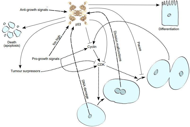
Indicate if each of the statements is true or false:A. Tumours which have lost p53 activity have higher mutation rates than tumours which have p53 activity.
B. Single celled organisms, such as yeast (Saccharomyces), possess equally powerful regulators like p53.
C. Treatments which deliver more p53 to cells, would reduce the division of healthy cells.
D. Both alleles of p53 are usually mutated in cancer. E. P53 knockout mice (Mus musculus) show an overgrowth of bone-marrow cells.
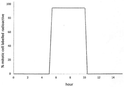
An experiment was set up to observe cell cycle length of a strain of yeast. Activated yeast cells were subcultured into a new medium with an initial concentration of 106 cells/mL. After 40 hours, the number of cells increased to 4 x 10⁶ cells/mL. A portion of the culture was taken for a separate experiment. In this experiment, cells were incubated for 15 min into a media containing radioactive thymidine before washing and re-grown on a new media containing non-radioactive thymidine. Cell samples were then taken periodically to measure the percentage of mitotic cells containing radioactive thymidine. The figure shows the result obtained from the experiment. At each sampling, about 1% of the total cells sampled were undergoing mitosis. Indicate if each of the following statements are True or False:A. G2 phase of the cell cycle takes approximately 10 hours.
B. Most of the yeast cells in the culture are at G1.
C. M phase of the cell cycle takes longer than 1 hour.
D. Most of the radioactive thymidine is assimilated in the S phase of the cell cycle.
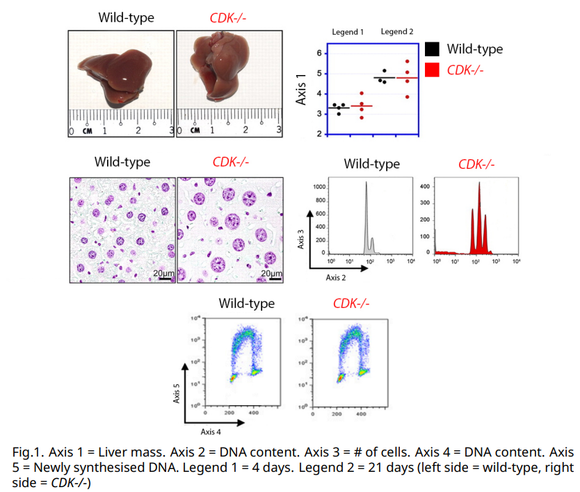
CDK is a family of cyclin dependent kinases which drive the cell cycle. A portion of the liver was removed in WT mice, or in livers which had a particular CDK gene knocked-out. Mice were allowed to regenerate, then their regenerated livers were weighed, measured and stained. Individual liver cells were removed and stained for total DNA content, or for newly synthesised DNA (Figure 1)
Indicate with an X whether you would expect number or amount increased (+), not changed (0) or decreased (-) of the following structures in CDK knock-out livers versus wild-type ones.
A. Liver massB. Hepatocyte numberC. Average hepatocyte ploidyD. Number of mitotic spindlesE. DNA synthesis
Practice Problems
1. A cell entering mitosis has a mutation that prevents kinetochore-microtubule attachments from being properly sensed by the spindle assembly checkpoint. Predict the most likely immediate consequence for the resulting daughter cells.
Show answer
Anaphase can proceed even with improperly or unattached kinetochores, risking unequal chromosome segregation (aneuploidy) in the resulting daughter cells: one daughter may receive an extra chromosome copy, the other may be missing one, since the checkpoint that would normally halt the cycle until every chromosome is correctly bi-oriented has been disabled.
2. A student states “meiosis I separates sister chromatids, and meiosis II separates homologous chromosomes.” Correct this statement, and explain the consequence of this mix-up for predicting genetic outcomes.
3. Explain, mechanistically, why plant cells cannot use an actin-myosin contractile ring for cytokinesis the way animal cells do, and describe the structural alternative plant cells use instead.