Nucleotide & Nucleic Acid Chemistry
Overview
This page covers nucleotide and nucleic acid chemistry from the biochemist’s angle: the bonds, functional groups, and chemical differences between DNA and RNA. DNA Structure & Replication builds directly on this to cover the double helix and replication machinery; that division mirrors how Lipids & Membrane Biochemistry covers phospholipid chemistry while Plasma Membrane Structure & Transport covers the resulting membrane’s behaviour.
Key Concepts
Nucleotide structure
A nucleotide has three components: a nitrogenous base, a five-carbon (pentose) sugar, and one or more phosphate groups. A nucleoside is the base-sugar unit alone, without phosphate. Naming convention: base + sugar + phosphate count determines the name (e.g. deoxyadenosine triphosphate, dATP: adenine + deoxyribose + 3 phosphates).
Purines and pyrimidines
Nitrogenous bases fall into two structural classes:
- Purines, double-ring structures (fused six- and five-membered rings): adenine (A) and guanine (G).
- Pyrimidines, single six-membered ring: cytosine (C), thymine (T), and uracil (U).
Each purine pairs specifically with one pyrimidine via hydrogen bonding, A with T (or U in RNA), G with C, a pairing rule that is chemically, not arbitrarily, determined: the hydrogen bond donor/acceptor pattern only matches correctly in the A–T/A–U and G–C combinations, and pairing two purines together would sterically clash (too wide) while pairing two pyrimidines would leave them too far apart to hydrogen-bond. G–C pairs form three hydrogen bonds, A–T pairs form two: which is why GC-rich DNA regions have a measurably higher melting temperature ($T_m$) than AT-rich regions of the same length.

The sugar: ribose vs. deoxyribose
RNA uses ribose; DNA uses 2′-deoxyribose: the single chemical difference is the absence of a hydroxyl group at the 2′ carbon in DNA. This is not a cosmetic difference: the 2′-OH in RNA makes the sugar-phosphate backbone chemically more reactive and prone to alkaline/enzymatic hydrolysis, meaning RNA is intrinsically less chemically stable than DNA, a major reason DNA, not RNA, is the long-term hereditary information store in cellular life, while RNA’s greater reactivity is co-opted functionally in ribozymes (catalytic RNA, whose 2′-OH groups can participate directly in phosphodiester bond chemistry).

Phosphodiester bonds and strand directionality
Nucleotides are joined by phosphodiester bonds: the 5′-phosphate of one nucleotide’s sugar links to the 3′-hydroxyl of the next, forming a sugar-phosphate backbone with a defined chemical direction: one end terminates in a free 5′-phosphate, the other in a free 3′-hydroxyl. This directionality (5′ → 3′) is not a labelling convention; it dictates the direction every polymerase can synthesise in (see DNA Structure & Replication and Transcription & RNA Processing for why this constraint shapes the replication fork and creates leading/lagging strand asymmetry).
DNA vs. RNA: the chemistry behind the functional divergence
| Feature | DNA | RNA | Functional consequence |
|---|---|---|---|
| Sugar | 2′-deoxyribose | Ribose | RNA more reactive/unstable, suited to transient messages, not long-term storage |
| Pyrimidine | Thymine (5-methyluracil) | Uracil | The extra methyl group in thymine is a repair signal (see below) |
| Strandedness | Almost always double-stranded | Usually single-stranded (can fold intramolecularly) | Single-strandedness lets RNA adopt complex catalytic/structural folds (tRNA cloverleaf, ribozymes) that duplex DNA cannot |
| Stability | High | Low | Matches DNA’s role as permanent archive vs. RNA’s role as disposable working copy |
Why thymine, not uracil, in DNA: cytosine spontaneously deaminates to uracil at a low but non-negligible rate. If DNA used uracil natively, the cell’s repair machinery could not distinguish “a uracil that belongs here” from “a uracil produced by cytosine deamination damage.” Using thymine (uracil + a methyl group) as DNA’s normal pyrimidine means any uracil found in DNA is unambiguously a deamination product, which base-excision repair (uracil-DNA glycosylase) can recognise and excise specifically, see DNA Repair & Recombination. This is one of the clearest examples in all of biochemistry of a chemical detail existing specifically to support an error-correction mechanism.
Comparative Structures
| Comparison axis | Purine (A, G) | Pyrimidine (C, T, U) |
|---|---|---|
| Ring structure | Fused 6+5 membered, two rings | Single 6-membered ring |
| Relative size | Larger | Smaller |
| H-bonds in canonical pairing | 2 (with T/U) or 3 (G with C) | Same, pairing-dependent |
| Pairs exclusively with | One specific pyrimidine | One specific purine |
Common Exam Questions
- “Why does GC content affect DNA melting temperature?”: always cite the hydrogen bond count difference (3 vs. 2), not simply “GC pairs are stronger” without the mechanistic reason.
- “Why does DNA use thymine instead of uracil?”, the repair-signal argument (distinguishing genuine thymine from deaminated-cytosine-derived uracil) is the complete, examinable answer; “thymine is more stable” alone is an incomplete answer.
- Questions describing an unfamiliar nucleic-acid-derived molecule (e.g. a modified nucleotide analog used as a drug) often hinge on identifying which chemical group (2′-OH presence/absence, base identity) has been altered and predicting the functional consequence by analogy to the DNA/RNA comparison above.
Visual Reference
Interactive
- A base-pairing hydrogen-bond viewer: click any two bases and see whether/how many hydrogen bonds form, reinforcing why only A–T/U and G–C pairings are chemically valid.
Static
(Static images are placed inline in Key Concepts above, next to the concept each one illustrates, rather than collected here. Two items are not yet illustrated: a nucleotide structure diagram, the sourced candidate for this slot, nucleotide-structure-diagram.jpg, turned out to duplicate the ribose-vs-deoxyribose comparison already placed above rather than showing a full base+sugar+phosphate nucleotide, so it was not inserted (see image-sources.md), and a phosphodiester backbone/5′→3′ directionality diagram, for which no image was sourced yet.)
Practice Challenge
Competition-sourced practice questions for this topic, graded by difficulty. Click the Solution tab to reveal each answer.
Nucleic acids, a group of macromolecules, are characterized by distinctive structures and functions. Match the phrases on the left with the appropriate word or phrase on the right. [NOTE: Answers may be used more than once]
| 1. Monomer of Nucleic Acids | a. Phosphate group |
|---|---|
| 2. A nucleotide is a sugar, a phosphate and a _________ | b. Deoxyribose |
| 3. Sugar in DNA | C. RNA |
| 4. Passed from parent to offspring | D. Nitrogenous base |
| 5. Overall structure of DNA | E. A, T, C, G |
| 6. Sugar of one nucleotide bonds to ________ of the next nucleotide | F. DNA |
| 7. DNA is expressed through this intermediary | G. Ribose |
| 8. Nitrogenous bases of DNA | H. Double Helix |
| 9. Nitrogenous bases of RNA | I. Nucleotide |
| 10. Sugar in RNA | J. A, U, C, G |
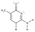
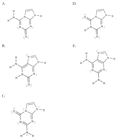
Watson-Crick base pairing relies on hydrogen bonding between nitrogenous bases. Chemists synthesized the artificial non-ACTG nitrogenous base shown on the right. Suppose an artificial strand of DNA is synthesized using this nitrogenous base. Which of the following artificial nitrogenous bases is MOST likely to pair with the nitrogenous base shown above via THREE hydrogen bonds?
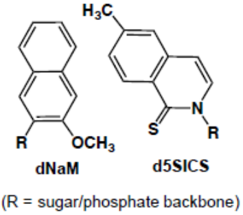
Organisms are defined by the information encoded in their genome, which can either be DNA or RNA. Essentially, there are four bases (A, T, G and C) that undergo specific complementary base pairing per Chargaff’s rule to form the DNA genome. Recently, two synthetic nucleotides were introduced into bacteria, which are designed to form a new complementary base pair of d5SICS-dNaM as shown below.
A. In a bacterial cell, how many possible tri-nucleotides (codons) exist using the bases A, T, G, C, d5SIC and dNaM?
B. Choose the most likely bonding (choose from covalent bond/hydrogen bond/ionic bond/ hydrophobic interaction/Van Der Waal forces) that allows d5SICS-dNaM base pairing and specify how it differs from the typical purine-pyrimidine base pairing.
Practice Problems
1. Two double-stranded DNA fragments of equal length have different GC content: Fragment A is 70% GC, Fragment B is 30% GC. Which has the higher melting temperature, and why?
Show answer
Fragment A (70% GC). G–C base pairs form three hydrogen bonds versus two for A–T pairs, so a GC-rich fragment requires more thermal energy to separate the strands: melting temperature increases with GC content.
2. A researcher finds an unusual uracil base incorporated into a sample of genomic DNA. Explain, mechanistically, how the cell’s repair machinery can recognise this as an error requiring correction, using the DNA/RNA base-composition rule.
3. Explain why RNA’s 2′-OH group, often described simply as making RNA “less stable” than DNA, is also the chemical basis for RNA’s ability to act as an enzyme (a ribozyme), i.e. why is the same feature both a stability liability and a functional asset?