Translation & the Genetic Code
Overview
Translation converts the nucleotide sequence of mature mRNA (see Transcription & RNA Processing) into the amino acid sequence of a polypeptide (see Amino Acids & Protein Chemistry Fundamentals for the chemistry of the product), read out through the genetic code. This page covers the code’s key properties, the adaptor molecule (tRNA) that physically links codon to amino acid, ribosome structure, and the three-stage translation mechanism.
Key Concepts
Properties of the genetic code
The genetic code maps 64 possible codons (three-nucleotide sequences) to 20 amino acids plus stop signals, with several defining properties, each with real biological consequences:
- Degenerate (redundant): most amino acids are specified by more than one codon (up to 6 for some), reducing but not eliminating the impact of single-nucleotide mutations: a mutation changing a codon to a synonymous codon (same amino acid) is a silent mutation with no protein-level consequence.
- Unambiguous: each codon specifies only one amino acid (or stop); there is no reverse ambiguity, even though the reverse mapping (amino acid → codon) is one-to-many.
- Nearly universal: the same codon-to-amino-acid mapping is used across nearly all known life (with a small number of documented exceptions, e.g. some mitochondrial genetic codes), strong evidence for a shared evolutionary origin of the translation system itself.
- Non-overlapping and read in a fixed frame: codons are read consecutively, three nucleotides at a time, without overlap, starting from a fixed reading frame set by the start codon. This is the direct reason insertions or deletions that are not multiples of three (frameshift mutations) are so much more disruptive than single-base substitutions: every downstream codon is shifted, typically scrambling the entire downstream amino acid sequence and frequently introducing a premature stop codon.

Open reading frames (ORFs)
An open reading frame (ORF) is a continuous stretch of codons, beginning with a start codon (AUG) and ending with a stop codon, uninterrupted by a stop codon in between: the sequence that, if translated, would produce a complete polypeptide. An ORF is a property of the nucleotide sequence (mRNA, or genomic DNA when scanned computationally, as in the ORF-finding tools covered in Bioinformatics); a protein’s primary structure (see Protein Structure, Folding & Function) is the actual linear amino acid sequence that results once that ORF is translated. The relationship is direct and one-directional under normal translation: a valid ORF, read through the genetic code, specifies a unique primary structure, but the ORF itself is nucleic acid information, while primary structure is the realised polypeptide product; the two are related exactly the way a recipe (ORF) is related to the finished dish (primary structure), not interchangeable terms for the same thing.
tRNA: the adaptor molecule
Transfer RNA (tRNA) physically links a specific codon to its corresponding amino acid, folding into a characteristic cloverleaf secondary structure (further folding into an L-shaped tertiary structure) with an anticodon loop at one end (base-pairing with the mRNA codon) and the amino-acid-attachment site at the other (the 3′ end). Aminoacyl-tRNA synthetases, one specific enzyme per amino acid, catalyse tRNA charging: covalently attaching the correct amino acid to its matching tRNA, consuming ATP in the process. This charging step is where the genetic code’s fidelity is actually enforced at the molecular level: the synthetase must correctly match both the amino acid and the tRNA’s anticodon identity; an error here (attaching the wrong amino acid to a tRNA) would cause the ribosome to insert an incorrect amino acid despite perfectly correct codon-anticodon pairing, since the ribosome itself has no independent way to verify that a charged tRNA carries the “correct” amino acid for its anticodon.
Wobble base pairing: the third codon position often tolerates non-standard base pairing with the anticodon’s first position, allowing a single tRNA species to recognise more than one synonymous codon: the direct molecular explanation for why the genetic code’s degeneracy is concentrated overwhelmingly at the third codon position.

Ribosome structure
The ribosome is a ribozyme-containing ribonucleoprotein complex (rRNA plus protein) with two subunits, small (reads and verifies codon-anticodon pairing) and large (contains the peptidyl transferase catalytic centre that forms peptide bonds, notably, this catalytic activity is carried out by rRNA itself, not a protein, making peptide bond formation a ribozyme-catalysed reaction), that assemble on the mRNA only during active translation. Three functional tRNA-binding sites span both subunits: the A (aminoacyl) site receives each incoming charged tRNA, the P (peptidyl) site holds the tRNA carrying the growing polypeptide chain, and the E (exit) site is where the now-uncharged tRNA sits briefly before release.

The three stages of translation
- Initiation: the small ribosomal subunit, together with an initiator tRNA (charged with methionine in eukaryotes), assembles at the mRNA’s start codon (AUG): in eukaryotes, this typically involves the small subunit binding near the 5′ cap (see Transcription & RNA Processing) and scanning downstream to locate the first AUG; the large subunit then joins to complete an assembled ribosome, with the initiator tRNA positioned in the P site.
- Elongation: a charged tRNA matching the next codon enters the A site; the ribosome catalyses peptide bond formation between the A-site amino acid and the growing chain held in the P site; the ribosome then translocates one codon along the mRNA, shifting the tRNAs from A→P and P→E, freeing the A site for the next incoming charged tRNA. This cycle repeats codon by codon.
- Termination: when a stop codon (UAA, UAG, UGA, none of which are recognised by any tRNA) enters the A site, a release factor binds instead, triggering hydrolysis of the completed polypeptide from the final tRNA and disassembly of the ribosomal complex.
Post-translational modification, revisited
The newly synthesised polypeptide is not necessarily functional immediately: folding (assisted by chaperones), and covalent modifications including phosphorylation, glycosylation (see Amino Acids & Protein Chemistry Fundamentals), and proteolytic cleavage of signal sequences or pro-peptide regions, are frequently required before the mature, functional protein is complete.
Comparative Structures
| Component | Role in translation | Key structural feature |
|---|---|---|
| mRNA | Carries the codon sequence (ORF) | Read 5′→3′, three nucleotides per codon |
| tRNA | Adaptor linking codon to amino acid | Cloverleaf/L-shaped fold; anticodon loop + amino-acid attachment site |
| Aminoacyl-tRNA synthetase | Charges tRNA with its correct amino acid | One enzyme per amino acid, the actual fidelity checkpoint |
| Ribosome (small subunit) | Codon-anticodon verification | Binds mRNA, scans for start codon |
| Ribosome (large subunit) | Peptide bond catalysis | Peptidyl transferase centre, RNA-catalysed (ribozyme) |
Common Exam Questions
- “Why is a frameshift mutation typically more disruptive than a point substitution?”: the correct answer must invoke the non-overlapping, fixed-reading-frame property of the code, every downstream codon changes, not just one.
- “Where is translation fidelity actually enforced, given the ribosome cannot check whether a charged tRNA carries the ‘right’ amino acid?”, tests understanding that fidelity is enforced upstream, at the aminoacyl-tRNA synthetase charging step, not at the ribosome itself.
- “What is the relationship between an ORF and a protein’s primary structure?”: the correct answer distinguishes the nucleic acid sequence property (ORF) from the resulting polypeptide (primary structure), rather than treating the two terms as synonyms.
- “Why does wobble pairing matter?”: explains why fewer than 61 distinct tRNA species can still read all 61 sense codons, concentrated at the third codon position.
Visual Reference
Interactive
- A ribosome translocation animator: step through initiation, then repeated elongation cycles (A-site charging, peptide bond formation, translocation), then termination, with the growing polypeptide chain visibly extending each cycle.
Static
(Static images are placed inline in Key Concepts above, next to the concept each one illustrates, rather than collected here.)
Practice Challenge
Competition-sourced practice questions for this topic, graded by difficulty. Click the Solution tab to reveal each answer.
Several codons of the mitochondrial DNA cytochrome b gene are shown.
| Columba junoniae | AAC GGG ATA CGT ACA GAT ATC ATT CCC CTA TAT CGG CCA CCC CTT ACA |
|---|---|
| Columba arquatrix | AAC AGG ATA CGT GCA AAT ATC ATT CCC CTA CAT CGG CCA TCC CTT ACA |
| Columba trocaz | GAC GGG ATA TGT ACA AAT ATC GTT CCC ATA CAT CGG CCA TCC CTT ACA |
| Columba palumbus | GAC AGG ATA TGT ACA AAT ATC GTT CCC ATA CAT CGG TCA CCC CTT ACA |
| Columba bolli | AAC GGG ATA TGT ACA AAT ATC GTT TCC ATA CAT CGG CCA TCC CTT GCA |
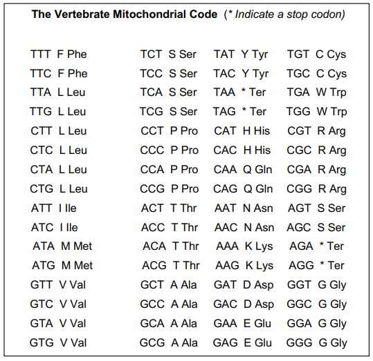
Based on DNA sequences and the Vertebrate mitochondrial DNA code, indicate which statements are true or false:
A. There are no amino-acid substitutions between species Columba junoniae and Columba bolli in the codons shown. B. There are two codon substitutions between species Columba trocaz and Columba palumbus in the codons shown. C. One amino-acid substitution from Columba arquatrix to Columba junoniae, in the codons shown, is from Ala to Thr. D. There are 8 amino-acid substitutions between species Columba junoniae and Columba bolli in the codons shown. E. There are 18 amino-acid substitutions between species Columba junoniae and Columba bolli in the codons shown.
A synthetic mRNA sequence 5′-AUG UUA GGU UGG UAA-3′ was translated in vitro using wild-type E. coli ribosomes and yielded a peptide Met-Leu-Gly-Trp. When the same mRNA was translated in a mutant E. coli strain, the product peptide was: Met-Leu-Gly-Trp-Tyr. All known tRNAs and aminoacyl-tRNA synthetases were the same as wild-type, 3 except one species of tRNA in the mutant carried tyrosine but had the anticodon AUU. Which interpretation/s best explain/s the altered translation outcome? i. The mutant tRNA reads the UAA stop codon, inserting tyrosine instead of terminating translation. ii. The mutant ribosome fails to recognize start codons, extending translation downstream. iii. A frameshift occurred at the glycine codon, producing an altered peptide tail with tyrosine near the C-terminus.
A. i onlyB . iii onlyC . i and iii onlyD . i, ii and iii
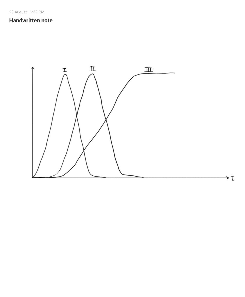
A) A researcher synchronizes the incorporation of a single amino acid into a growing polypeptide and continuously monitors three covalent linkages associated with that amino acid. The following graph shows the results of fluorescent probing of 3 different bonds involved in translation.
Match the correct bond to the number (I, II or III). Options for bonds are: Acid-anhydride (phospho-anhydride) bond, ester bond and peptide bond.
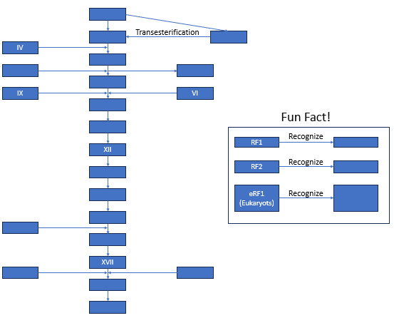
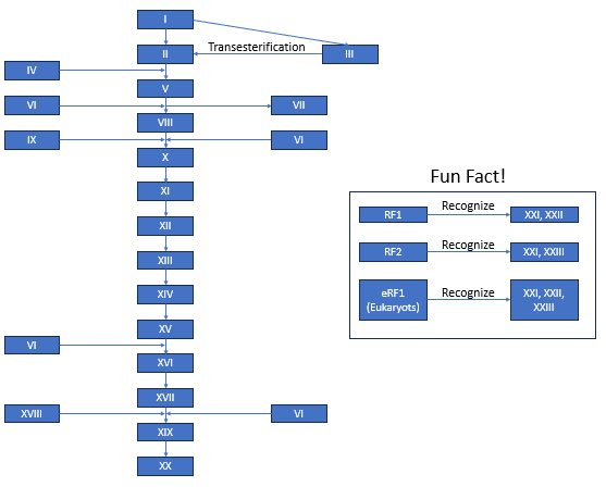
B) One day while visiting the library, you find the librarian very distressed. Upon further inquiry, you get to know that a book issued by her is now missing a page containing the solution to the following flowchart explaining the steps of translation in E. coli. Being IBO aspirants, you now need to fill the flowchart with the correct alternatives (one may be repeated many times). You are given following alternatives:
Attachment of Amino Acid to ATP by formation of acid-anhydride (phospho-anhydride) bond and release of Pyrophosphate
Amino Acid is transferred to 3’OH of A residue at 3’ end of tRNA
Amino Acid is transferred to 2’OH of A residue at 3’ end of tRNA
Binding of Aminoacyl-tRNA to GTP bound EF-Tu complex
Aminoacyl-tRNA-EF-Tu-GTP complex binds to A site of ribosome
Hydrolysis of GTP
Release of EF-Tu-GDP complex
Formation of peptide bond between Amino Acid in P site and A site
Formation of EF-G-GTP complex
Translocation of ribosome 1 codon towards 3’ end of mRNA
tRNA of P site reaches E site and is released into cytoplasm
Termination codon reaches A site
tRNA mimicking RF1/RF2 occupy A site and growing polypeptide is transferred to H2O causing the polypeptide to leave the ribosome
RF3-GDP binds to ribosome to cause release of RF1/RF2
GTP replaces GDP in RF3-GDP
RF3 is released from ribosome
tRNA mimicking Ribosome Recycling Factor (RRF) binds to A site of Ribosome.
Formation of EF-G-GTP complex
Translocation or ribosome causing RRF to move to P site and uncharged tRNA (corresponding to codon preceding stop codon) to move to E site
RRF causes releases of - c tRNA and EF-G releases RRF to cause all subunits to dissociate
UAA
UAG
UGA
Practice Problems
1. What is the difference between an open reading frame and a protein’s primary structure, and what is the relationship between the two?
Show answer
An open reading frame (ORF) is a property of a nucleotide sequence: a continuous stretch of codons from a start codon to an in-frame stop codon, uninterrupted by any stop codon in between. Primary structure is the resulting linear amino acid sequence of the actual translated polypeptide. The relationship is that of a template to its product: translating a valid ORF through the genetic code directly specifies a unique primary structure, but the two are not the same kind of object, an ORF is nucleic acid sequence information, while primary structure is the realised polypeptide chain that results from decoding it.
2. A single-nucleotide substitution changes a codon from GAA (Glu) to GAG. Predict the effect on the resulting protein, and name this category of mutation.
3. A mutation in an aminoacyl-tRNA synthetase gene causes it to occasionally charge its designated tRNA with an incorrect amino acid, while codon-anticodon recognition at the ribosome remains completely normal. Explain why this error would go completely undetected by the ribosome’s own proofreading mechanisms, and predict the consequence for the resulting protein.