Carbohydrate Chemistry & Biology
Overview
Carbohydrates are built from one simple repeating chemical idea, the glycosidic bond, yet that single linkage, varied in stereochemistry and branching pattern, produces molecules as different as the rigid, indigestible cellulose of a plant cell wall and the readily mobilised glucose reserve of glycogen. This page covers monosaccharide chemistry, how glycosidic bonds form and are named, and why storage/structural polysaccharides differ so sharply in physical properties despite near-identical monomer composition.
Key Concepts
Monosaccharide stereochemistry
Monosaccharides are polyhydroxy aldehydes (aldoses, e.g. glucose) or ketones (ketoses, e.g. fructose). Each chiral carbon doubles the number of possible stereoisomers, so glucose (4 chiral centres in its open-chain form) has 16 possible stereoisomers, of which D-glucose is the biologically overwhelming default.
D/L designation is assigned by the configuration at the chiral carbon farthest from the carbonyl group (compared to D/L-glyceraldehyde as the reference); nearly all biologically relevant sugars are D-sugars, mirroring the near-universal use of L-amino acids in proteins; both conventions reflect that the enzymes handling these molecules are themselves chiral and only accept one stereoisomeric family efficiently.
In aqueous solution, 5- and 6-carbon sugars predominantly exist as cyclic hemiacetals/hemiketals (furanose or pyranose rings), not open chains. Ring closure creates a new chiral centre at C1 (the anomeric carbon), giving rise to α and β anomers, e.g. α-D-glucopyranose vs. β-D-glucopyranose, which interconvert in solution (mutarotation) but are locked into one form once incorporated into a glycosidic bond. This α/β distinction is the single most consequential fact in this page: it is the entire reason starch/glycogen and cellulose behave so differently (see below).

Glycosidic bonds
A glycosidic bond forms between the anomeric carbon of one sugar and a hydroxyl group of another, releasing water (condensation): the carbohydrate equivalent of a peptide bond. Bonds are named by the two carbons joined and the anomeric configuration, e.g. α(1→4) links C1 of one glucose to C4 of the next in an α configuration.
Storage vs. structural polysaccharides: the same monomer, opposite function
Starch (plants) and glycogen (animals) are both glucose polymers linked almost entirely by α(1→4) bonds, with α(1→6) branch points (more frequent in glycogen than in amylopectin, giving glycogen a more densely branched, “bushier” structure with more non-reducing ends for rapid simultaneous mobilisation by glycogen phosphorylase). The α linkage puts a consistent kink at every bond, so the chain naturally coils into a helix, a shape enzymes (amylase, phosphorylase) can access easily from the outside, which is exactly what a rapidly mobilised energy store needs.


Cellulose, by contrast, links glucose exclusively by β(1→4) bonds. The β linkage forces each successive glucose to rotate 180° relative to its neighbour, producing a straight, extended chain rather than a helix. These straight chains pack side-by-side into extensive inter-chain hydrogen-bonded sheets (microfibrils), which is what gives cellulose its tensile strength and near-total resistance to digestion by animal enzymes (which recognise only α-glycosidic geometry; only specialised cellulase-producing organisms, often microbial symbionts, can hydrolyse β(1→4) bonds).
This is the highest-yield single comparison in carbohydrate biochemistry: identical monomer, only the anomeric configuration of the linkage differs, and that alone explains storage-vs-structural function.

Glycosaminoglycans and the extracellular matrix
Beyond glucose homopolymers, glycosaminoglycans (GAGs), long, unbranched, highly negatively charged polymers of repeating disaccharide units (e.g. hyaluronic acid, chondroitin sulfate, heparin), are a distinct structural carbohydrate class central to the extracellular matrix (see Cell Junctions, Extracellular Matrix & Cell Death). Their dense negative charge draws in water and cations, giving connective tissue its resistance to compression.
Glycosylation, revisited
Amino Acids & Protein Chemistry Fundamentals introduced N- and O-glycosylation as protein modifications; from the carbohydrate side, the attached glycan itself is built by sequential glycosyltransferase reactions in the ER and Golgi, each adding one sugar via a new glycosidic bond, and each enzyme recognising both the specific sugar donor and the specific acceptor hydroxyl. This stepwise, enzyme-templated (rather than nucleic-acid-templated) assembly is why glycan structures are far more heterogeneous between individual protein molecules than a directly gene-encoded sequence like a polypeptide chain.
Comparative Structures
| Polysaccharide | Monomer linkage | Chain shape | Branching | Biological role |
|---|---|---|---|---|
| Starch (amylose) | α(1→4) | Helical | None | Plant energy storage |
| Starch (amylopectin) | α(1→4), α(1→6) branches | Helical with branches | Moderate | Plant energy storage |
| Glycogen | α(1→4), α(1→6) branches | Helical with branches | Dense (more than amylopectin) | Animal energy storage, rapid mobilisation |
| Cellulose | β(1→4) | Extended, straight | None | Plant cell wall structure |
| Chitin | β(1→4) of N-acetylglucosamine | Extended, straight | None | Fungal cell wall, arthropod exoskeleton structure |
Common Exam Questions
- “Why can’t humans digest cellulose?”: the correct answer names the β(1→4) linkage geometry, not simply “we lack the enzyme,” since the follow-up (“why don’t we have that enzyme?”) is really asking about substrate specificity for α- vs. β-glycosidases.
- “Compare glycogen and amylopectin structurally”: degree of branching (glycogen denser) is the testable distinction, since both use the same α(1→4)/α(1→6) linkage chemistry.
- Anomeric carbon identification (which carbon is C1, why ring closure creates a new stereocentre there) is a frequent structure-drawing question.
Visual Reference
Interactive
- An α vs. β glycosidic bond builder: click two monosaccharide ring diagrams together in either configuration and see the resulting chain shape (helical vs. extended) render automatically, making the single-cause structural argument of this page directly visible.
Static
(Static images are placed inline in Key Concepts above, next to the concept each one illustrates, rather than collected here.)
Practice Challenge
Competition-sourced practice questions for this topic, graded by difficulty. Click the Solution tab to reveal each answer.
Read the following observations and some interpretations. Observations:
- To preserve the sweet taste of a freshly picked corn, it is immersed in boiling water for a few minutes.
- The sweetness of honey decreases if it is heated.
- Commercial fructose cannot be used as a sweetener for hot drinks.
Interpretations: I. The properties of sugar vary with temperature. II. Enzyme that converts starch to glucose is activated due to rise in temperature. III. Some sugars are thermostable hence their properties do not change with change in temperature. IV. Enzyme that converts glucose to starch is destroyed due to rise in temperature.
Mark the correct interpretation against each observation.
1 - _____________2 - _____________3 -
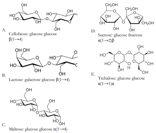
The primary disaccharide digestion product of starch is
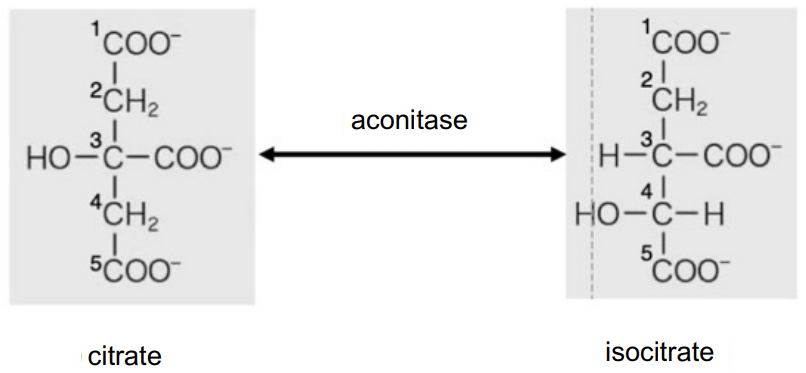
The citric acid cycle is central to metabolism, for the supply energy and various key compounds. In citric acid cycle, the enzyme aconitase catalyzes the reversible conversion between citrate and isocitrate. In this reaction, OH group at C3 and H group at C4 of citrate are removed as water, thereafter a water molecule is added back in a reverse manner to generate isocitrate as given in figure. However, OH group is never added at C2.
Indicate whether each of the following statements is True or False: A. Citrate has enantiomers B. Isocitrate has enantiomers C. Two:CH2COO- groups are stereochemically equivalent when citrate is free in solution D. Two:CH2COO- groups are stereochemically equivalent when citrate is bound to aconitase
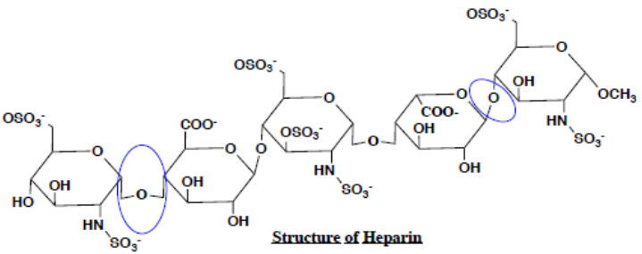
The following is the structure of heparin, which is naturally produced by basophils and mast cells in the body and is also manufactured and distributed as the most widely used anticoagulant.
A. Which class of macromolecules best describes heparin: (lipids/nucleic acids/carbohydrates/ proteins)
B. Identify the circled covalent bonds as glycosidic/peptide/phosphodiester/ester bond and list the byproduct of the reaction that results in the formation of this covalent bond.
C. Heparin is often used to purify nucleic acid (both DNA and RNA) binding proteins. Explain what the similarity between heparin and nucleic acids is that allows them to bind to these proteins.
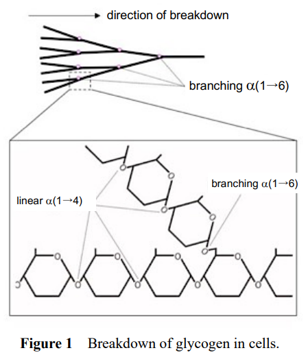
Glycogen (and amylopectin) is a glucose polymer with some branching. Linear chains of these polymers consist of α(1→4) linkages and occasional branching is formed by α(1→6) linkage (Figure 1). For degradation in cells, glucose residues are released one-by-one from the end of the chains by phosphorylase up to the residue at the branching site. Then, the α(1→6) branching site is removed by a debranching enzyme.
- Given that a certain glycogen consisting of 10000 glucose residues is branched at every 10 residues, how many terminal chains are available for phosphorylase?
A. A bout 10B. A bout 50C. About 100D. About 500E. A bout 1000F. About 5000
- For degradation of this glycogen by excess phosphorylase or by excess debranching enzyme, choose an appropriate graph for its breakdown from below. Assume that the phosphorylase releases all glucose residues from a linear chain without branching.
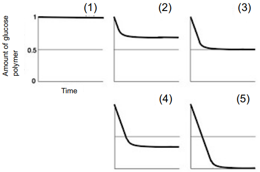
- Plant amylopectin is similar to glycogen but branching occurs much less frequently. Given that the branching of an amylopectin of similar size of glycogen is formed at every 25 residues, indicate the combination of correct descriptions about degradation of amylopectin by phosphorylase.
A. Breakdown speed is slower than that of glycogenB. Breakdown speed is similar to that of glycogenC. Breakdown speed is faster than that of glycogenD. Final breakdown extent is smaller than that of glycogenE. Final breakdown extent is similar to that of glycogenF. Final breakdown extent is larger than that of glycogen
Practice Problems
1. A polysaccharide is found to be completely resistant to human salivary amylase but readily digested by a specific bacterial enzyme. Propose the most likely glycosidic linkage type, and justify your reasoning.
Show answer
Most likely a β-glycosidic linkage (e.g. β(1→4), as in cellulose). Human amylase is specific for α-glycosidic bonds; resistance to it but susceptibility to a specialised bacterial enzyme (a cellulase-type glycosidase) is the classic signature of β-linked polysaccharides, which only organisms with the appropriate β-glycosidase machinery can hydrolyse.
2. Explain, at the level of glycosidic bond geometry, why glycogen is well-suited to be rapidly mobilised for energy but cellulose could never function as a usable energy reserve for the organism that makes it (independent of enzyme availability).
3. If a mutation eliminated all α(1→6) branch points from glycogen synthesis (converting it to a linear α(1→4) chain only, like amylose), predict the effect on the rate of glucose mobilisation from a glycogen granule of the same total mass, and explain why.