Human Digestive System
Overview
This page covers the GI tract from mouth to anus and its accessory organs (liver, gallbladder, pancreas) at full structural depth, region by region, since each region’s histology is specifically adapted to a distinct digestive task.
Key Concepts
GI Tract Wall: General Plan
Every region of the tract shares four concentric layers, innermost to outermost, though the tissue within each layer changes region to region: mucosa (epithelium in direct contact with contents, plus a thin underlying connective tissue layer and a thin smooth muscle layer, the muscularis mucosae), submucosa (connective tissue, blood vessels, and the submucosal (Meissner’s) nerve plexus), muscularis externa (usually inner circular + outer longitudinal smooth muscle layers, with the myenteric (Auerbach’s) nerve plexus between them, coordinating peristalsis), and serosa (outer connective tissue, continuous with the peritoneum in the abdominal cavity). Both nerve plexuses are part of the enteric nervous system, capable of coordinating digestive motility semi-independently of CNS input, a structurally embedded, not merely centrally controlled, nervous network.
Oral Cavity
Teeth are anchored in sockets (alveoli) of the maxilla/mandible by the periodontal ligament. Each tooth has a crown (exposed, capped by enamel, the hardest substance in the body, acellular and avascular, unable to regenerate if damaged) and a root (embedded in bone, covered by cementum rather than enamel); beneath enamel/cementum, dentin (a living, mineralized tissue, softer than enamel) forms the bulk of the tooth, surrounding a central pulp cavity (blood vessels, nerves, connective tissue). Adult humans have 32 teeth, heterodont (see Mammalian Comparative Anatomy for the broader mammalian pattern): incisors (cutting), canines (tearing), premolars and molars (grinding).

Three paired salivary glands deliver secretions via ducts into the oral cavity: parotid (largest, anterior to the ear, serous secretion, high enzyme content), submandibular (mixed serous/mucous), sublingual (mostly mucous). Saliva begins starch digestion (via salivary amylase) and lubricates food for swallowing. The tongue (skeletal muscle, see Human Muscular System) manipulates food and bears taste buds clustered on papillae.
Esophagus and Swallowing
The esophagus is a muscular tube with no digestive/absorptive function, purely conductive, histologically distinguished by stratified squamous epithelium (abrasion-resistant, appropriate for transiting solid food, see Body Plans) and, uniquely among GI regions, a muscularis externa that transitions from skeletal muscle (upper third, allowing voluntary initiation of swallowing) to smooth muscle (lower two-thirds, involuntary). Two functional (not strictly anatomically distinct) sphincters bound it: the upper esophageal sphincter (relaxes during swallowing, otherwise prevents air entry) and the lower esophageal sphincter (LES) (relaxes to admit food into the stomach, otherwise prevents reflux of acidic gastric contents; LES dysfunction is the structural basis of reflux disease, a useful applied reference point).
Stomach
Grossly divided into the cardia (adjacent to the LES), fundus (superior dome), body (largest region), and pylorus (leading to the pyloric sphincter, regulating emptying into the duodenum); the internal mucosa is thrown into folds, rugae, which flatten as the stomach distends, allowing substantial volume expansion. The mucosa is densely populated with gastric pits, each leading to gastric glands containing several specialized secretory cell types, a high-yield histology table:
| Cell type | Location | Secretion | Function |
|---|---|---|---|
| Mucous neck cells | Upper gland/pit | Mucus (alkaline) | Protects epithelium from acid |
| Parietal cells | Mid-gland | HCl, intrinsic factor | Acidifies lumen (activates pepsinogen, kills pathogens); intrinsic factor is required for vitamin B12 absorption later in the ileum |
| Chief cells | Base of gland | Pepsinogen (inactive) | Converted to active pepsin by the acidic environment HCl creates: a structural safeguard against the stomach digesting its own protein-based secretory cells |
| G cells | Base of gland (pyloric region) | Gastrin (hormone, into blood, not the lumen) | Stimulates parietal cell HCl secretion |

🧪 Gastric Gland Cell-Type Matcher
The stomach epithelium itself is protected from its own acidic, protein-digesting contents by a thick alkaline mucus layer and rapid epithelial cell turnover, a structural, not just chemical, defense.
Small Intestine
Three regions with distinguishing structural features, worth naming individually rather than treating “small intestine” as uniform: duodenum (shortest, receives bile and pancreatic secretions via the hepatopancreatic ampulla, contains Brunner’s glands in the submucosa, unique to this region, secreting alkaline mucus to neutralize acidic chyme arriving from the stomach), jejunum (middle, the site of most nutrient absorption, has the most prominent villi and plicae circulares), ileum (longest, contains Peyer’s patches, aggregated lymphoid nodules, an immune surveillance structure specific to this region, since the ileum is the last checkpoint before the microbially dense large intestine, and is the specific site of vitamin B12 and bile salt reabsorption).
Absorptive surface amplification, structurally layered: plicae circulares (circular mucosal/submucosal folds, visible grossly) → villi (finger-like mucosal projections, each containing a capillary network for nutrient absorption and a central lymphatic lacteal for fat absorption) → microvilli (brush border on individual absorptive epithelial cells). Between villi, the mucosa dips into crypts of Lieberkühn, containing proliferative stem cells (replenishing the rapidly turned-over epithelium) and secretory cells.
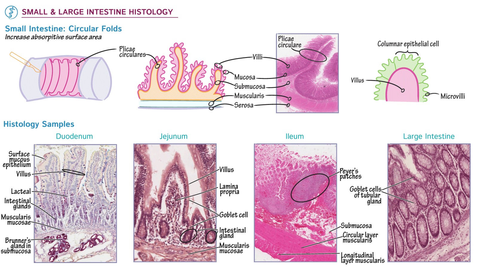 Source: (originally via ditki.com). Excellent match, directly shows each region’s unique distinguishing marker (Brunner’s glands, Peyer’s patch) named in the text, side by side with the absorptive-hierarchy diagram.

Large Intestine
Begins at the cecum (a blind pouch, bearing the vermiform appendix, lymphoid tissue of debated but likely immune/microbiome-reservoir function), continuing through the ascending, transverse, descending, and sigmoid colon to the rectum and anal canal (bearing an internal involuntary smooth-muscle sphincter and an external voluntary skeletal-muscle sphincter, a structural pairing directly analogous to the LES/pyloric-sphincter pattern of involuntary control, but here paired with a voluntary override). Structurally distinct from the small intestine: no villi (absorptive surface reduced, since the primary remaining job is water/electrolyte reabsorption and waste consolidation, not nutrient absorption), and the longitudinal muscle layer is gathered into three thickened bands, the teniae coli, whose tonic contraction gathers the wall into pouches, haustra, a distinctive gross-anatomical signature of the colon on imaging or a specimen. The colon houses a dense resident microbial population (the gut microbiome), structurally supported by this large, slow-transit surface, contributing to fermentation of otherwise-undigested material and synthesis of some vitamins.

Liver
The functional and structural unit is the hepatic lobule: a roughly hexagonal arrangement of hepatocyte plates radiating from a central vein, with a portal triad (hepatic artery branch, hepatic portal vein branch, bile duct branch) at each of the hexagon’s corners. Blood from both the hepatic artery (oxygenated, ~25% of liver blood flow) and the hepatic portal vein (nutrient-rich, deoxygenated, draining the GI tract, ~75% of flow, meaning nearly everything absorbed by the small intestine passes through the liver before reaching general circulation) mixes and flows through sinusoids (large, fenestrated capillary-like channels, see Human Circulatory System) past hepatocyte plates toward the central vein. This is the reverse direction to bile flow, which moves from hepatocytes into small bile canaliculi between cells, then outward to the bile duct at the portal triad. Kupffer cells (specialized resident macrophages) line the sinusoids, filtering the blood of pathogens/debris, a direct immune function tied to the liver’s position downstream of the entire GI tract’s absorbed contents.

Gallbladder and Pancreas
The gallbladder, a small muscular sac beneath the liver, stores and concentrates bile (produced continuously by the liver) between meals, releasing it via the cystic duct (joining the common hepatic duct to form the common bile duct) into the duodenum when stimulated. The pancreas has a dual structural identity: the vast majority of its tissue is exocrine, organized into grape-like clusters (acini) of enzyme-secreting cells draining via a duct system into the pancreatic duct, which joins the common bile duct at the hepatopancreatic ampulla; scattered through this exocrine tissue are small, discrete islets of Langerhans, endocrine tissue (structurally distinct clusters, not connected to the duct system, secreting hormones (insulin, glucagon) directly into the bloodstream) that make up under 2% of pancreatic mass but perform a completely separate function from the surrounding exocrine acini.

Peritoneum
The abdominal cavity is lined by the parietal peritoneum (body wall) and covers most abdominal organs with visceral peritoneum, structurally analogous to the pleura around the lungs (see Human Respiratory System). Double-layered peritoneal folds, mesenteries, suspend the intestines from the posterior body wall while carrying their blood vessels, nerves, and lymphatics; the greater omentum (a large, fat-laden peritoneal fold hanging from the stomach, draping over the intestines) and lesser omentum (connecting the stomach/duodenum to the liver) are named peritoneal extensions relevant to gross anatomical/surgical orientation.
🍽️ GI Transit Walkthrough
Click a stop directly, or use Next/Previous to trace a bite of food from mouth to anus, assembling every region's histology, secretions, and transit time into one connected path.
Comparative Structures
The four-layer GI tract wall plan, and the accessory-gland (liver/pancreas) arrangement, are broadly conserved across vertebrates, with gut length and regional specialization tracking diet, see the Vertebrate Anatomy tier pages and, for the ruminant four-chambered stomach as a specific herbivore adaptation, Mammalian Comparative Anatomy. Invertebrate guts show a simpler version of the same regional-specialization principle (e.g. the earthworm crop/gizzard) without the liver/pancreas-level accessory gland complexity, see Invertebrate Body Plans I.
Common Exam Questions
- “Name the four gastric gland cell types and, for each, state its secretion and explain how the structural arrangement (chief cells secreting an inactive zymogen) prevents self-digestion.”
- “Distinguish the duodenum, jejunum, and ileum by one unique structural feature each.”
- “Explain the functional significance of the hepatic portal vein’s course, in terms of what the liver is structurally positioned to do to absorbed nutrients before they reach systemic circulation.”
- “A patient’s gallbladder is removed. Explain, structurally, what function is lost, and why bile itself can still reach the duodenum.”
- “Explain why the pancreas is described as having a dual (exocrine/endocrine) structural identity, referencing the acini and islets specifically.”
Visual Reference
Interactive
(Implemented inline above: the gastric gland cell-type matcher quiz sits directly below the gastric gland image, and the GI transit walkthrough sits at the end of Key Concepts, after the Peritoneum section.)
Static
(Static images are placed inline in Key Concepts above, next to the concept each one illustrates, rather than collected here.)
Practice Problems
- Trace a bite of food from the mouth to the point where it first encounters bile, naming every organ/region it passes through in order.
- Explain why chief cells secrete pepsinogen rather than active pepsin directly, and what converts it to its active form.
- A structural feature unique to the ileum aids both immune surveillance and vitamin B12 absorption timing. Name it and explain its regional placement.
- Compare the direction of blood flow to the direction of bile flow within a hepatic lobule.
- Explain why the islets of Langerhans are described as functionally and structurally separate from the exocrine pancreas despite being embedded within it.