Fish & Amphibian Anatomy
Overview
This is the first of the three Vertebrate Anatomy pages, and every comparison on it is made explicitly against the human structures covered in the Human Anatomy tier. Fish and amphibians are grouped together because amphibians are structurally transitional between fish and fully terrestrial vertebrates. An amphibian’s anatomy only makes sense as “a fish body plan partway adapted to land,” which is also why amphibian development (aquatic larva → terrestrial adult, most visible in frogs) recapitulates that transition within a single life cycle.
Key Concepts
Fish Skeleton and Fins
Two major skeletal grades: cartilaginous (Chondrichthyes: sharks, rays; skeleton is cartilage throughout, never ossifies to bone, though it may be calcified for added rigidity) and bony (Osteichthyes, the vast majority of fish species; ossified skeleton, plus a swim bladder, a gas-filled dorsal organ regulating buoyancy by adjusting gas volume; developmentally related to the structures that become lungs in tetrapods, a direct homology worth stating explicitly rather than treating the two organs as unrelated). Fins are supported by fin rays (bony or cartilaginous rods, not the tetrapod limb-bone pattern); paired pectoral and pelvic fins provide steering and stabilization, while the unpaired dorsal, anal, and caudal fins provide additional stability and, at the caudal fin, primary thrust via lateral body/tail undulation.
Fish Respiratory Structure
Gills, not lungs: each gill arch bears a row of primary lamellae (filaments), and each primary lamella bears many thinner secondary lamellae projecting from its surface, the true site of gas exchange, each covered in a single layer of epithelium directly apposed to a dense capillary bed (the same “minimal diffusion distance” principle as the human alveolus, see Human Respiratory System). Water flows across the secondary lamellae in the direction opposite to blood flow within them (countercurrent exchange), which maintains a favorable diffusion gradient along the entire length of the lamella (unlike concurrent flow, where the gradient would collapse once blood and water oxygen levels equalized partway across), allowing bony fish to extract a much higher fraction of dissolved O₂ from water than concurrent flow would permit.

🐟 Countercurrent vs. Concurrent Gill Exchange
In bony fish, all four gill arches on each side are covered by a bony flap, the operculum, whose rhythmic movement (paired with mouth opening/closing) actively pumps water across the gills even when the fish is stationary; cartilaginous fish generally lack an operculum and rely more on forward swimming (ram ventilation) or dedicated spiracles.

Fish Circulatory Structure
A two-chambered heart (one atrium, one ventricle, preceded by a thin-walled collecting chamber, the sinus venosus, and followed by an outflow tract, the conus arteriosus or bulbus arteriosus depending on the group) and a single circuit: blood is pumped once, passes through the gills to pick up oxygen, then continues directly to the body before returning to the heart via the sinus venosus. Compare this to the human double-circuit, four-chambered heart on the Human Circulatory System page. A fish heart only ever pumps blood that is about to lose pressure crossing the gill capillary bed before it reaches the body, a structural limitation double circulation solves.

Fish Osmoregulatory Structure
Fish kidney structure differs by habitat in a way directly comparable to the nephron detail on the Human Excretory System page: freshwater fish (hypertonic to their environment, constantly gaining water by osmosis) have kidneys with large, numerous glomeruli producing copious dilute urine; marine bony fish (hypotonic to seawater, constantly losing water) have reduced or in some species absent glomeruli (aglomerular nephrons), filtering far less and instead relying heavily on active secretion at the tubule and on specialized gill chloride cells to actively excrete excess salt, a clear structural inversion between the two habitats’ nephron populations.

Fish Sensory Structure: The Lateral Line
A structure with no direct human equivalent: the lateral line system, a row of fluid-filled canals running along each side of the body and over the head, opening to the surrounding water through pores. Within the canals, clusters of mechanoreceptor hair cells (neuromasts, structurally similar in principle to the cochlear/vestibular hair cells on the Human Sensory Organs page) are deflected by water movement/pressure changes, allowing fish to detect nearby movement, obstacles, and prey, a distributed mechanosensory structure supplementing the eyes and inner ear.
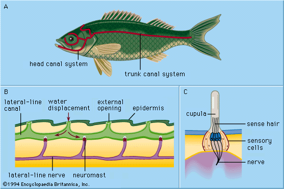 Source: Encyclopædia Britannica, Inc.
Amphibian Skeleton
The first vertebrates with true tetrapod limbs (the proximal-bone/distal-bones/digits pattern directly comparable to the human arm/leg plan, see Human Skeletal System), though weaker and more sprawling than in reptiles/mammals, with the limbs positioned lateral to the body (requiring more muscular effort to support body weight against gravity than the more vertically positioned mammalian limb). Amphibians still rely partly on water’s buoyancy to support body weight efficiently, which is why most remain tied to aquatic or moist habitats.
Amphibian Respiratory Structure
A genuine structural hybrid, often using three surfaces simultaneously: gills (larval stage, e.g. tadpoles, lost at metamorphosis in most species, a direct structural echo of the fish gill plan above), simple saclike lungs (much less internally divided than mammalian lungs, far less surface area, a direct structural explanation for why amphibians can’t sustain the metabolic rate of birds/mammals), and cutaneous respiration (direct gas exchange across thin, moist, minimally keratinized, highly vascularized skin, meaning amphibian skin must stay permeable and moist, the structural reason amphibians are so vulnerable to desiccation and skin-absorbed toxins). Amphibians additionally lack a diaphragm (unlike the mammalian mechanism on the Human Respiratory System page) and instead ventilate their lungs by positive-pressure buccal pumping: floor-of-mouth movements force air into the lungs, structurally the reverse mechanical strategy to the negative-pressure suction ventilation used by mammals.

Amphibian Circulatory Structure
A three-chambered heart (two atria, one ventricle): a structural improvement on the fish heart (separate return paths from the body and the lungs/skin into two atria) but an incomplete one, since both atria still empty into a single, undivided ventricle. Mixing is reduced below what the shared chamber alone would suggest, however, by two structural features worth naming specifically: the ventricle’s internal wall is trabeculated (ridged, spongy), which helps maintain some separation of oxygenated and deoxygenated blood streams as they pass through by favoring laminar, minimally mixing flow; and the spiral valve, a ridge within the conus arteriosus/outflow tract, preferentially directs the first (more deoxygenated) blood leaving the ventricle toward the pulmonary/cutaneous circuit and later (more oxygenated) blood toward the systemic circuit. This is a direct, structurally specific example of “functional separation without complete anatomical separation” and a good exam topic precisely because it shows structure compensating, imperfectly, for what a full septum would otherwise provide.
🫀 Heart Blood-Flow Animator
Amphibian Skin
Thin, glandular, permeable (no scales in most adult amphibians, unlike fish or reptiles), the same epidermis-over-dermis tissue plan as human skin (see Human Integumentary System) but with minimal keratinization, dense mucous glands (keeping the surface moist, supporting cutaneous respiration) and, in many species, dense granular (poison) glands, a direct structural trade: a barrier permeable enough for gas exchange is also a barrier that offers little protection against desiccation or toxin absorption, which is why chemical defense (toxin-secreting glands) is so widespread in this lineage specifically.
Comparative Structures
| Feature | Fish | Amphibian | Human baseline (for reference) |
|---|---|---|---|
| Heart chambers | 2 (1 atrium, 1 ventricle) + sinus venosus | 3 (2 atria, 1 ventricle) | 4 (2 atria, 2 ventricles) |
| Circuit(s) | Single | Double, incompletely separated (trabeculae + spiral valve reduce mixing) | Double, fully separated |
| Gas exchange organ | Gills (countercurrent, lamellar) | Gills (larval) / saclike lungs / skin | Alveolar lungs (tidal) only |
| Ventilation mechanism | Opercular pumping / ram ventilation | Positive-pressure buccal pumping | Negative-pressure diaphragm/intercostal |
| Limb support | Fins (fin rays) | Tetrapod limbs (weak/sprawling, lateral) | Tetrapod limbs (upright) |
| Skin | Dermal bony scales, mucus-covered | Moist, permeable, glandular, minimal keratin | Keratinized, relatively impermeable |
| Unique sensory structure | Lateral line (neuromasts) | Reduced/lost in most adults | , |
Common Exam Questions
- “Explain why countercurrent flow across fish gill lamellae extracts more oxygen than concurrent flow would, referencing the diffusion gradient along the lamella’s length.”
- “An amphibian’s ventricle is anatomically undivided, yet blood entering the systemic and pulmonary circuits is not fully mixed. Explain the two structural features responsible.”
- “Compare the ventilation mechanism of a frog to that of a human, identifying which is a positive-pressure and which is a negative-pressure system.”
- “A marine bony fish has reduced or absent renal glomeruli compared to a freshwater relative. Explain this structural difference in terms of each fish’s osmotic environment.”
- “Describe the structure and function of the lateral line system, and explain why it has no equivalent in terrestrial vertebrates.”
Visual Reference
Interactive
(Implemented inline above: the countercurrent vs. concurrent gill exchange graph sits directly below the gill lamellae image, and the heart blood-flow animator sits directly below the amphibian circulatory structure paragraph.)
Static
(Static images are placed inline in Key Concepts above, next to the concept each one illustrates, rather than collected here.)
Practice Problems
- Trace a red blood cell through a fish’s single circuit, starting at the sinus venosus, and explain why blood pressure delivered to the body is lower than in a human.
- Explain the specific structural mechanism (naming both features) by which an amphibian heart limits mixing of oxygenated and deoxygenated blood despite lacking a ventricular septum.
- List the three respiratory surfaces available to an adult amphibian and state which is likely dominant when the animal is submerged versus on land.
- Explain why fish scales and amphibian skin represent two different anatomical solutions to living in water, despite both being aquatic-adapted, referencing tissue origin (dermal vs. epidermal).
- Explain the functional significance of the swim bladder being developmentally related to tetrapod lungs.