Invertebrate Body Plans II
Overview
This page continues directly from Invertebrate Body Plans I, covering three eucoelomate, triploblastic, bilaterally symmetric phyla (see Body Plans): Mollusca, Arthropoda, and Echinodermata. Mollusca and Arthropoda are protostomes, developmentally grouped with Annelida from the previous page; Echinodermata is a deuterostome, developmentally closer to Chordata (and therefore to every phylum in the Vertebrate Anatomy tier) despite looking, superficially, like the most “alien” body plan in this section.
Key Concepts
Mollusca
The second-largest animal phylum by species count, structurally unified by three body regions present (in some form) across all classes: the muscular foot (locomotion, a flat crawling sole in snails, siphon-based jet propulsion in cephalopods), the visceral mass (containing most internal organs), and the mantle (a tissue layer secreting the calcium carbonate shell, where present, and enclosing the mantle cavity, which houses the ctenidia: comb-like gills, each a central axis bearing rows of thin filaments across which water is drawn by ciliary action, the same “thin epithelium over a dense capillary-like network” gas-exchange logic seen throughout this section).

Structural diversity within the phylum is a major exam topic:
- Gastropods (snails, slugs): a single coiled shell (or none, in slugs/nudibranchs) and a unique feeding structure, the radula, a chitinous, tooth-covered ribbon drawn back and forth over a cartilage-like support, the odontophore, to scrape or shred food; found nowhere outside Mollusca. Gastropods also undergo torsion during larval development: the visceral mass and mantle cavity rotate up to 180° relative to the head/foot, bringing the mantle cavity (and anus) to a position above/near the head, a structurally counterintuitive developmental event (unrelated to shell coiling, a common student confusion) whose adaptive explanation remains a genuinely debated topic, making it a good higher-order exam question rather than a simple recall fact.

- Bivalves (clams, mussels, oysters): two hinged shells (valves) enclosing a laterally compressed body; no radula (bivalves are filter feeders, using enlarged ctenidia for both respiration and food capture, structurally repurposing the same gill tissue for two functions) and drawing/expelling water through two siphons (incurrent and excurrent), an unmistakable structural feature of an otherwise foot- and head-reduced body plan.
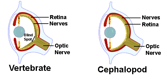
- Cephalopods (squid, octopus): the most structurally derived class: foot modified into arms/tentacles (each often bearing suckers), shell reduced/internal/absent, and by far the most complex invertebrate nervous system on this page, a large, centralized brain (formed from fused ganglia, encased in a cartilaginous cranium structurally analogous in function, though not origin, to the vertebrate skull) and, in squid, giant axons enabling extremely fast escape reflexes. Cephalopods have a closed circulatory system (blood confined entirely to vessels, plus two accessory “branchial hearts” pumping blood specifically through each gill), unlike the open system in most other mollusks, supporting their unusually high, fish-comparable metabolic activity. The cephalopod eye is a striking case of convergent evolution with the vertebrate camera eye (see Human Sensory Organs): independently evolved cornea, lens, iris, and a light-sensitive retina-equivalent layer, arranged in a strikingly similar overall geometry despite the two eye types being wired in opposite orientation (the cephalopod retina is not inverted the way the vertebrate retina is, meaning cephalopods have no structural blind spot equivalent).
 Source:
Chromatophores (pigment-filled cells under direct neural/muscular control, able to expand or contract almost instantly) in the skin give many cephalopods rapid, structurally active (not just chemical/hormonal) color change and camouflage capability.
Most mollusks (bivalves, gastropods) have an open circulatory system: the heart pumps blood (hemolymph) into open sinuses (a hemocoel) where it bathes tissues directly rather than staying confined to vessels, then diffuses back toward the heart, structurally simpler, and delivering oxygen/nutrients less efficiently, than the closed vertebrate system on the Human Circulatory System page.
Arthropoda
The largest animal phylum by a wide margin, defined by three linked structural features: jointed appendages, an exoskeleton, and segmentation with tagmatization. The phylum is structurally organized into four major subphyla, each with distinguishing features worth naming: Chelicerata (spiders, scorpions, horseshoe crabs: no antennae; anterior appendages are chelicerae rather than mandibles; body divided into cephalothorax and abdomen), Myriapoda (centipedes, millipedes: one pair of antennae; many similar leg-bearing trunk segments, minimal tagmatization compared to insects/crustaceans), Crustacea (crabs, lobsters, shrimp: two pairs of antennae, a structural feature unique to this subphylum; mostly aquatic, gill-bearing), Hexapoda (insects, three pairs of walking legs on a distinct thorax, one pair of antennae, most species winged).
| Subphylum | Antennae | Mouthpart-adjacent appendage | Body regions (tagmata) | Examples |
|---|---|---|---|---|
| Chelicerata | None | Chelicerae (not mandibles) | Cephalothorax + abdomen | Spiders, scorpions, horseshoe crabs |
| Myriapoda | 1 pair | Mandibles | Head + many similar trunk segments | Centipedes, millipedes |
| Crustacea | 2 pairs (unique to this subphylum) | Mandibles | Cephalothorax + abdomen | Crabs, lobsters, shrimp |
| Hexapoda | 1 pair | Mandibles | Head + thorax (3 leg-bearing segments) + abdomen | Insects |
🔑 Arthropod Subphylum Identification Key
- Exoskeleton: a rigid external covering made of chitin (a structural polysaccharide) hardened with proteins (and, in crustaceans, calcium carbonate). Muscle attaches to the inside of the exoskeleton rather than the outside of an internal skeleton, the opposite strategy to the vertebrate endoskeleton (see Human Skeletal System). The exoskeleton cannot grow, so arthropods must periodically molt (ecdysis), triggered hormonally (by ecdysone). The old exoskeleton is partially digested/detached from a newly secreted, temporarily soft cuticle beneath it, then shed, with the animal rapidly expanding (often by taking up water or air) before the new cuticle hardens; a vulnerable period exploited by predators.
- Segmentation with tagmatization: like Annelida, arthropods are fundamentally segmented, but segments are fused into specialized functional regions (tagmata): the three-part insect body (head/thorax/abdomen) or the two-part arachnid body (cephalothorax/abdomen) are both tagmatized versions of the ancestral fully-segmented arthropod plan.
Compound eyes, present in most crustaceans and insects, are a structurally distinct visual solution from the single-lens camera eye of vertebrates and cephalopods: each eye is built from many independent units, ommatidia, each with its own lens and photoreceptor cluster, together producing a mosaic image with a wide field of view and excellent motion detection, at the cost of lower spatial resolution than a single large lens/retina system can achieve, a direct structure-function contrast worth drawing against the vertebrate eye on the Human Sensory Organs page.

Like most mollusks, arthropods have an open circulatory system (hemolymph, hemocoel, a simple dorsal tubular heart with paired lateral openings, ostia, through which hemolymph re-enters the heart between contractions), but arthropod respiration bypasses the circulatory system for gas transport entirely in terrestrial forms: insects use a tracheal system, a network of branching tubes (tracheae, opening to the outside via spiracles, further subdividing into fluid-tipped tracheoles that deliver oxygen directly to individual tissue cells) with no need for blood-mediated gas transport at all, part of why insect body size is physically constrained (diffusion through tracheae becomes limiting at larger sizes). Insect excretion uses Malpighian tubules: blind-ended tubules projecting from the gut into the hemocoel, actively transporting waste (uric acid, structurally and chemically analogous to the reptilian/avian excretory strategy on the Reptile & Bird Anatomy page, both are terrestrial, water-conserving solutions arrived at independently) from the surrounding hemolymph into the gut lumen for elimination alongside feces. Aquatic crustaceans instead use gills, structurally convergent with (though not homologous to) fish gills and molluscan ctenidia.
A labeled diagram of the tracheal system and Malpighian tubules is already covered on the Comparative Osmoregulation & Excretion page in the Animal Physiology section, not repeated here.
Echinodermata
Deuterostomes (see Body Plans), and therefore developmentally the closest invertebrate phylum on this page to the Vertebrate Anatomy tier, despite the most anatomically distinctive body plan covered anywhere in this section. Adults show secondary radial symmetry (typically pentaradial, five-part) built on a bilaterally symmetric larval stage. The defining structural feature is the water vascular system: seawater enters through a sieve-like plate, the madreporite, passes down a stone canal to a circular ring canal, which distributes it into five (or a multiple of five) radial canals, each running the length of an arm and supplying a row of tube feet, each tube foot paired internally with a muscular, bulb-like ampulla; ampulla contraction forces fluid into the tube foot, extending it, while foot-muscle contraction retracts it, and a suckered tip provides adhesion, an entirely hydraulic, valve-and-muscle locomotor/feeding system with no structural equivalent anywhere else in the animal kingdom, used for locomotion, feeding (prying open bivalve shells in sea stars, notably including the mollusks covered earlier on this page), and gas exchange.

⭐ Water Vascular System Flow Animator
Echinoderms also have a calcareous endoskeleton: internal, mesoderm-derived ossicles embedded in the body wall (often bearing spines, the phylum’s namesake), a genuine endoskeleton structurally analogous in position (internal, living tissue covered by epidermis) to the vertebrate endoskeleton, despite evolving completely independently. Many species (notably sea stars) additionally bear tiny pincer-like pedicellariae on the body surface, used to keep the skin free of debris and small organisms. Gas exchange occurs via papulae (thin skin projections, “skin gills”) in sea stars, or via specialized structures in other classes, supplementing tube-foot exchange. Major classes: Asteroidea (sea stars), Ophiuroidea (brittle stars, more sharply demarcated arms than sea stars), Echinoidea (sea urchins/sand dollars, no arms, fused test/shell, long movable spines), Holothuroidea (sea cucumbers, elongated body, reduced skeleton, tube feet reduced/modified), Crinoidea (sea lilies/feather stars, often stalked, considered the most structurally ancestral class). Echinoderms also show pronounced regenerative capacity, some sea stars can regrow an entire arm, or in a few species even a whole body from a single arm with a fragment of the central disc, a structural/developmental parallel to the sponge archaeocyte regenerative capacity from the previous page, though arising through entirely different cellular mechanisms.
Comparative Structures
| Feature | Mollusca | Arthropoda | Echinodermata |
|---|---|---|---|
| Development | Protostome | Protostome | Deuterostome |
| Skeleton | External shell (where present), not a true exoskeleton | True chitinous exoskeleton, molted | Internal calcareous endoskeleton (ossicles) |
| Circulatory system | Open (most classes) / closed (cephalopods) | Open | No discrete circulatory system (water vascular system instead) |
| Gas exchange | Ctenidia (mantle cavity) | Tracheae (terrestrial) / gills (aquatic) | Tube feet / papulae |
| Locomotion structure | Muscular foot | Jointed appendages | Water-vascular tube feet |
| Nervous system | Ganglia (simple) to large centralized brain (cephalopods) | Ganglia, ventral nerve cord | Nerve ring + radial nerves, no brain |
| Excretory structure | Nephridia (metanephridia-like) | Malpighian tubules (terrestrial) / green glands (some crustaceans) | No discrete organ (diffusion via tube feet/papulae) |
Common Exam Questions
- “Explain why cephalopods, despite being mollusks, have a closed circulatory system unlike most other members of the phylum, and connect this to their metabolic demands.”
- “Compare the cephalopod eye and the vertebrate eye as an example of convergent evolution, identifying one structural similarity and one structural difference.”
- “A specimen has a rigid, jointed external covering made of chitin. Identify the phylum and explain the specific structural problem this covering creates for growth.”
- “Compare insect Malpighian tubules to reptilian/avian kidneys as independently evolved solutions to the same excretory problem.”
- “Trace the path of seawater through the water vascular system from the madreporite to a single extended tube foot.”
- “Justify the classification of Echinodermata as deuterostomes using developmental (not adult anatomical) evidence, and name the other major deuterostome phylum covered in this section.”
Visual Reference
Interactive
(Implemented inline above: the water vascular system flow animator sits directly below the water vascular system image, and the arthropod subphylum identification key sits directly below the subphylum comparison table.)
Static
(Static images are placed inline in Key Concepts above, next to the concept each one illustrates, rather than collected here. The arthropod subphyla comparison is a table in the text rather than an image; the insect tracheal system/Malpighian tubules are illustrated on the existing Animal Physiology page, cross-linked above rather than duplicated here.)
Practice Problems
- Name the three body regions shared across molluscan classes and briefly state how each is modified in cephalopods compared to gastropods.
- Explain why an arthropod must molt periodically, connecting your answer to the material composition of the exoskeleton, and name the hormone that triggers this process.
- A deuterostome invertebrate has secondary radial symmetry as an adult. Name the phylum and explain what “secondary” signals about its larval stage.
- Insects rely on a tracheal system rather than blood for gas transport. Explain why this places a physical upper limit on insect body size.
- Compare a compound eye and a camera-type eye by structural organization and explain the resulting trade-off between field of view and image resolution.