Endomembrane System & Protein Trafficking
Overview
The endomembrane system is a set of physically and functionally connected organelles, nuclear envelope, ER, Golgi apparatus, lysosomes, and the plasma membrane itself, linked by a continuous flow of membrane-bound transport vesicles rather than by direct physical continuity throughout. This page traces a secreted or membrane-bound protein’s journey from synthesis to final destination, covering each organelle’s role in sequence.
Key Concepts
Rough endoplasmic reticulum: the entry point
The rough ER is studded with ribosomes actively translating proteins destined for secretion, insertion into a membrane, or delivery into an organelle (versus cytoplasmic proteins, translated on free ribosomes and released directly into the cytosol). A signal sequence on the growing polypeptide is recognised by the signal recognition particle (SRP), which halts translation and directs the ribosome-mRNA complex to a translocon channel on the ER membrane; translation resumes, threading the growing chain directly into the ER lumen (for secreted/luminal proteins) or into the membrane itself (for transmembrane proteins, which stop threading at hydrophobic transmembrane-domain sequences). Inside the ER lumen, proteins undergo initial folding (assisted by ER-resident chaperones, see Protein Structure, Folding & Function) and N-linked glycosylation begins (see Amino Acids & Protein Chemistry Fundamentals and Carbohydrate Chemistry & Biology for the chemistry).
Smooth endoplasmic reticulum: synthesis and detoxification
The smooth ER lacks ribosomes and specialises in lipid synthesis (phospholipids, steroids), carbohydrate metabolism, and, particularly abundant in liver cells, detoxification of lipophilic compounds and drugs via cytochrome P450 enzymes. In skeletal muscle, a specialised smooth ER derivative (the sarcoplasmic reticulum) stores and releases Ca²⁺ to trigger contraction.
Quality control: ERAD
Proteins that fail to fold correctly in the ER are recognised by ER quality-control machinery and retrotranslocated back into the cytosol for degradation via ER-associated degradation (ERAD), which feeds into the ubiquitin-proteasome system covered in Protein Structure, Folding & Function. This is the exact mechanism underlying cystic fibrosis in most patients: the ΔF508 CFTR mutant folds incorrectly, is recognised by ERAD, and is degraded before ever reaching the plasma membrane, even though the mutant channel, if it did reach the membrane, would retain substantial function.
The Golgi apparatus: sorting, modifying, and shipping
Vesicles bud from the ER and fuse with the cis face of the Golgi apparatus, a stack of flattened membrane cisternae. As cargo moves through the stack toward the trans face, Golgi-resident enzymes further modify N-linked glycans, add O-linked glycosylation, and perform other covalent modifications in a spatially organised, assembly-line fashion, different enzymes are concentrated in different cisternae, so a protein’s modification state depends on how far through the stack it has travelled.

Lysosomes: degradation
Lysosomes are membrane-bound organelles containing acid hydrolases (proteases, nucleases, lipases, glycosidases) that function optimally at the lysosome’s maintained internal pH (~4.5–5), well below cytoplasmic pH: a protective feature, since accidental lysosomal membrane rupture releases enzymes that are far less active at neutral cytoplasmic pH, limiting collateral damage. They degrade material delivered via phagocytosis (engulfed particles/pathogens), autophagy (a double-membrane autophagosome engulfing the cell’s own damaged organelles or protein aggregates, which then fuses with a lysosome, see Cell Junctions, Extracellular Matrix & Cell Death for autophagy’s role in cell death pathways), and endocytosis (see below). Lysosomal storage diseases (e.g. Tay-Sachs) result from a deficiency in one specific acid hydrolase, causing its substrate to accumulate undegraded.

Peroxisomes: oxidation reactions, independent of the vesicular pathway
Peroxisomes are single-membrane organelles that perform oxidative reactions (notably very-long-chain fatty acid β-oxidation, which in animal cells occurs in peroxisomes rather than mitochondria for these particular substrates) and generate hydrogen peroxide as a byproduct, immediately neutralised by peroxisomal catalase. Unlike ER/Golgi/lysosome cargo, peroxisomal proteins are synthesised on free cytosolic ribosomes and imported post-translationally via a distinct signal-sequence-and-receptor system (PTS1/PTS2 signals), and peroxisomes replicate by growth and fission rather than budding from the ER/Golgi pathway, worth knowing as the clear exception to the “everything in the endomembrane system flows through vesicular budding” generalisation.
Vesicular trafficking: the connective tissue of the whole system
Endocytosis (phagocytosis, pinocytosis, receptor-mediated endocytosis) internalises extracellular material by inward budding of the plasma membrane; exocytosis is the reverse, vesicles fusing with the plasma membrane to release contents extracellularly or insert membrane proteins. Both directions rely on the same core molecular logic: coat proteins (e.g. clathrin, COPI, COPII) shape budding vesicles and select cargo, and SNARE proteins on the vesicle and target membrane pair specifically to ensure a vesicle fuses only with its correct destination membrane, this specificity is what keeps the whole multi-organelle trafficking system from collapsing into indiscriminate membrane mixing.

Comparative Structures
| Organelle/pathway | Ribosomes present? | Key function | Distinguishing feature |
|---|---|---|---|
| Rough ER | Yes | Co-translational folding, N-glycosylation start | Entry point for secretory/membrane proteins |
| Smooth ER | No | Lipid synthesis, detoxification, Ca²⁺ storage | No ribosomes; abundant in liver, muscle |
| Golgi apparatus | No | Further glycosylation, sorting | Cis-to-trans directional modification and sorting |
| Lysosome | No | Degradation (acid hydrolases) | Low internal pH; receives endocytic/autophagic/Golgi-derived cargo |
| Peroxisome | No | Oxidative reactions, VLCFA β-oxidation | Post-translational import; replicates by fission, not budding |
Common Exam Questions
- “Trace the path of a secreted protein from synthesis to release”: the expected answer sequence is rough ER (co-translational entry, initial folding/glycosylation) → Golgi (further modification, sorting) → secretory vesicle → plasma membrane (exocytosis); questions often ask you to identify which single step is missing or altered given an experimental perturbation.
- “Why does a misfolded CFTR protein cause disease even though the underlying channel, if present at the membrane, would work?”, tests understanding of ERAD as a quality-control checkpoint that can be a disease mechanism in itself, independent of the protein’s intrinsic function.
- Distinguishing peroxisomes from lysosomes is a common confusion point: both are single-membrane, roughly similar-sized organelles, but one degrades (lysosome, acidic, endomembrane-vesicle-derived) and one oxidises (peroxisome, near-neutral, post-translationally imported).
Visual Reference
Interactive
- A protein-trafficking pathway tracer: click a starting point (ribosome) and watch an animated vesicle carry a cargo protein sequentially through rough ER → Golgi → final destination, with each stage’s modification labelled as it happens.
Static
(Static images are placed inline in Key Concepts above, next to the concept each one illustrates, rather than collected here.)
Practice Challenge
Competition-sourced practice questions for this topic, graded by difficulty. Click the Solution tab to reveal each answer.
To trace the path of a secretory protein from its synthesis to its export from a cell, suppose that you have added radioactive amino acids to a culture of cells, and then measured the amount of radioactivity that shows up in the proteins of each of the following cell fractions at different times after addition. I: Secretory Vesicles II: Golgi Complex III: Rough Endoplasmic Reticulum IV: Nucleus
A. III -> II -> IV -> I -> out of the cell B. III -> II -> I ——–> out of the cellC. IV -> III -> II -> I -> out of the cellD. IV -> II -> III-> I -> out of the cell
Indicate if each of the following statements about posttranslational modification of eukaryotic proteins is true or false:A. Disulfide bond formation on a protein occurs in the endoplasmic reticulum.
B. Glycoproteins may be found in viruses that infect humans.
C. Oligosaccharide group addition to a protein may occur in both Golgi apparatus and endoplasmic reticulum.
D. Palmitoylation of a protein can change its intracellular localization.
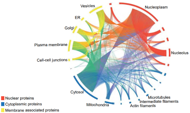
The location of proteins in a cell affects their function by determining which molecules they interact with. A collaboration of scientists in Sweden and Cambridge used microscopy to map the location of >12 000 human proteins in numerous cell types. In the diagram below, solid bars around the circumference represent all the different types of protein within each organelle, and lines are drawn between the same type of protein in different organelles.Indicate if each of the following statements is true or false:A. Most proteins are found only in a single organelle.
B. Most mitochondrial proteins are found only in the mitochondria.
C. Most proteins that are found in multiple organelles are spread homogeneously through the cell.
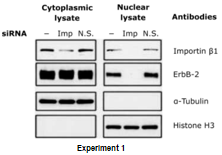
ErbB-2 is a receptor found on the plasma membrane of mammalian cells which can move from the plasma membrane to the nucleus. Because most proteins that shuttle between the cytoplasm and the nucleus are soluble and not integral membrane bound proteins, the mechanism by which ErbB-2 undergoes transport to the nucleus is of particular interest. Three experiments described below were done to shed light on the underlying mechanisms. Experiment 1: siRNA knock down of importin β1 expression in target cells. The cells were transfected with importin β1 siRNA (Imp), non-functional siRNA control (N.S.), or buffer only (−). Proteins from the cytoplasmic and nuclear fraction of cell lysates were analysed by Western blotting using importinβ1, and ErbB-2, α tubulin and histone H3 antibodies as indicated.
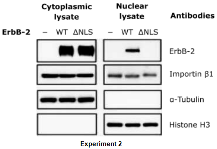
Experiment 2: Mutant cells lacking ErbB-2 gene were transfected with wild-type ErbB-2 (WT), ErbB-2 mutant containing a deficient nuclear localization signal (∆NLS), or vector control (−). Proteins from the cytoplasmic fraction, nuclear fraction were then analysed by Western blotting with ErbB-2, importinβ1, α tubulin and histone H3 antibodies as indicated.
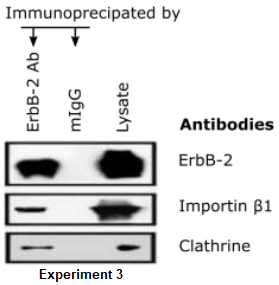
Experiment 3: Cell lysates from the cells were immune-precipitated with anti-ErbB-2 or mouse IgG (mIgG). The precipitated immune-complexes and the cell lysates were then analysed by Western blotting with, clathrin, importinβ1, and ErbB-2 antibodies.
Indicate if each of the following statements is true or false:A. The data in Experiment 1 suggest that ErbB-2 requires importin β1 in order to enter the nucleus.
B. It is predicted that the antibody against importin β1does not precipitate the ErbB-2(∆NLS).
C. The data presented suggest that localization of histones to the nucleus is mediated by a mechanism distinct from that used to shuttle ErbB-2.
D. As true for other membrane bound receptors, ErbB2 enters the cytoplasm by endocytosis.
Practice Problems
1. A mutation eliminates the mannose-6-phosphate tagging system in a cell line. Predict what happens to newly synthesised lysosomal enzymes, and to the cell’s ability to degrade endocytosed material.
Show answer
Without the mannose-6-phosphate tag, the Golgi cannot sort these enzymes into the lysosome-bound vesicle pathway; they are instead default-routed via constitutive secretion and released from the cell. Lysosomes become progressively deficient in acid hydrolases, and the cell’s ability to degrade endocytosed or autophagic material declines, functionally resembling a lysosomal storage disease, even though the enzymes themselves are made correctly and are catalytically normal once outside the cell.
2. Explain why lysosomal rupture is comparatively less damaging to a cell than an equivalent rupture of, say, a compartment containing highly active enzymes at neutral pH would be, referencing the lysosome’s internal chemical environment.
3. A drug blocks SNARE-mediated membrane fusion specifically. Predict the effect on both the secretory pathway (Golgi → plasma membrane) and the endocytic pathway (plasma membrane → early endosome → lysosome), and explain why this single mechanistic target affects both directions of vesicular traffic.